HOME > 写真 > 花・植物 > 葉
10,000件の写真素材が検索されました。
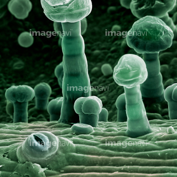
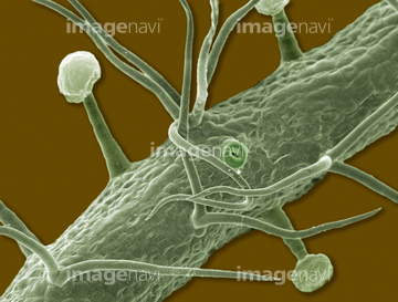
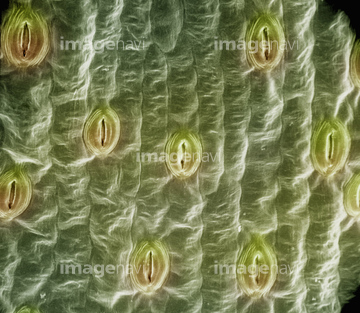
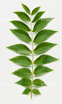
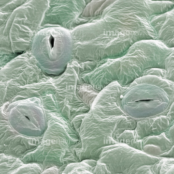
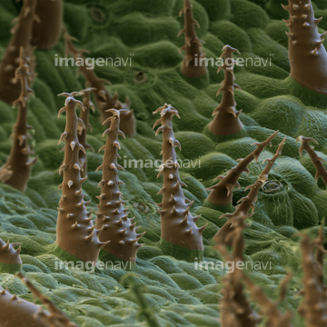
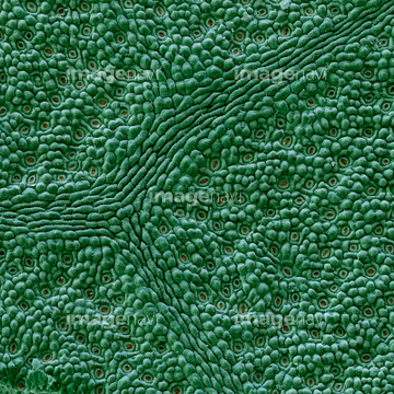
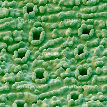
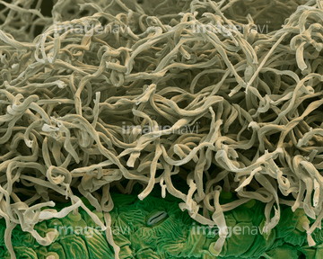
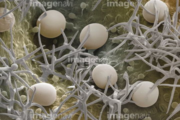
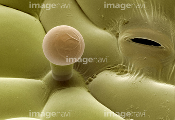
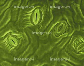
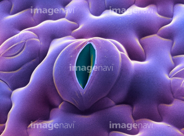
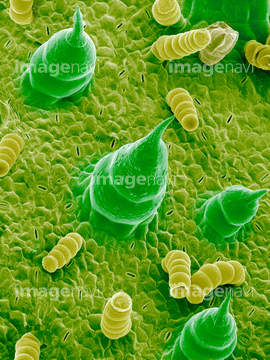
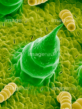
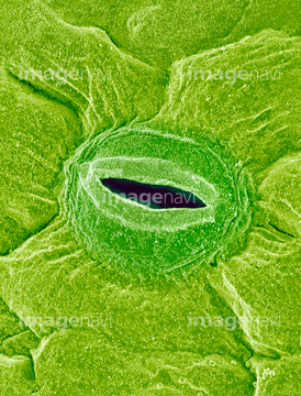
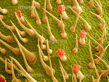
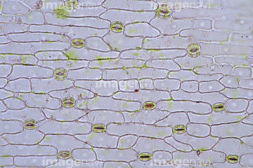
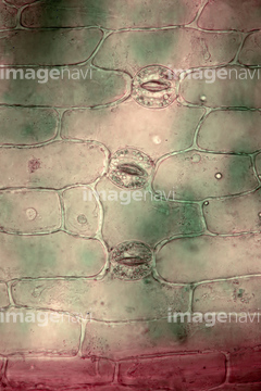
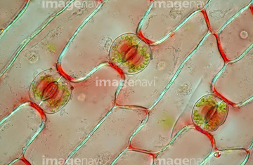
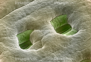
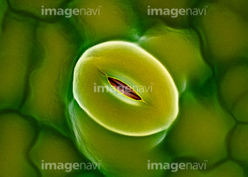
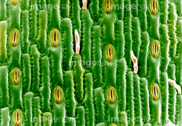
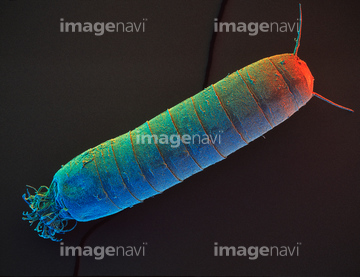
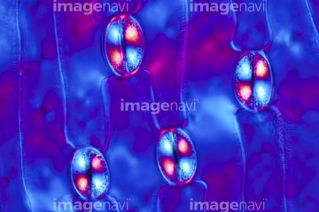
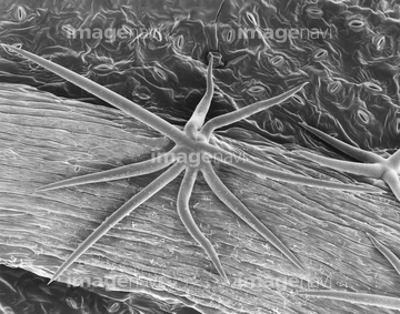
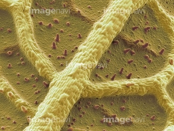
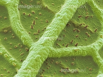
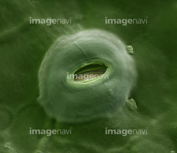
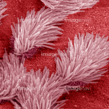
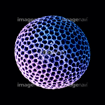
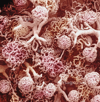
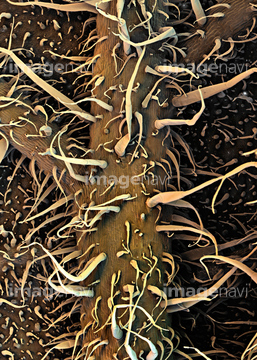
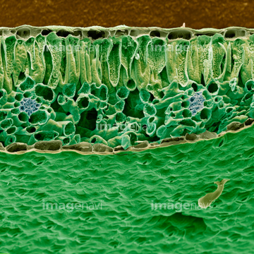
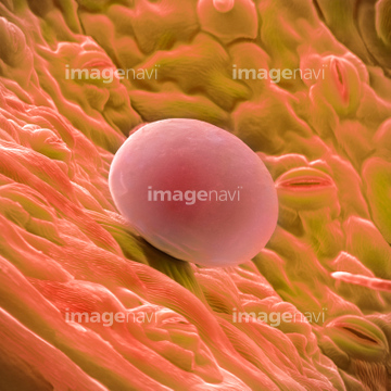
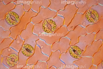
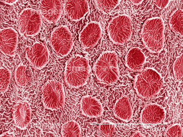
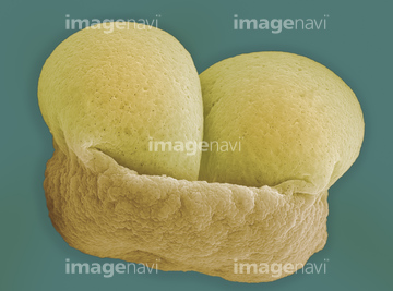
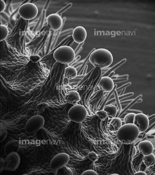
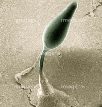
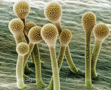
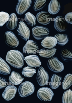
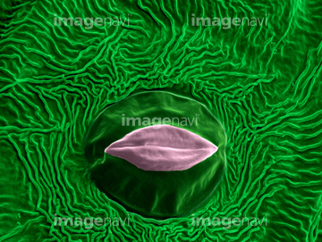
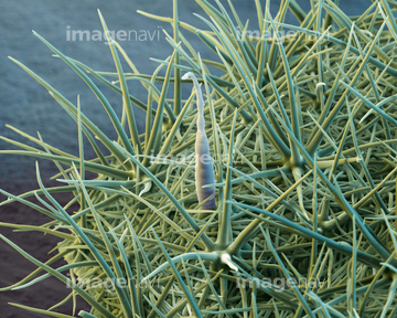
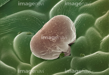
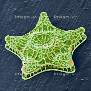
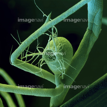
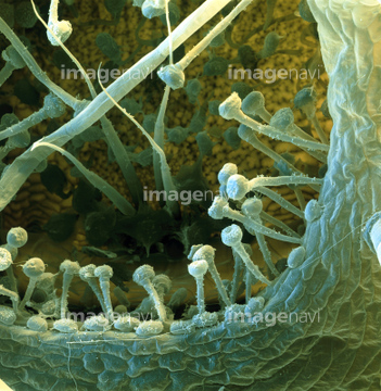
この検索結果には、Haworthia leaf、Witch-hazel leaf surface, SEM、Daisy leaf surface, SEM、Closed stoma, SEM、Open stoma, SEM、Sunflower leaf, SEMなどが含まれています。
64114726
64115526


64114192
64239426
64005629
64005630
64005632
64005633
64005638
64005643
64259882
64259884
64259885
64259886
64259887
64259888
64259889
64259890
64259891
64148624
64005622
64005631
64005642
64005644
64005648
64005657
64005665
64005666
64005675
64005688
64005689
64005709
64005765
64005766

64004044
64005627
64005628
64005637
64005681
64005682
64192019
20542488
64255079
64255080
64255901
64157866
64056494
64005641
64005683
64005684
64005699
64005703
64005706
64005716
64005717
64005722
64005761
64005762
64005763
64004043
64089408
64148626
64005390
64006294
64006295
64006296
64005704
64005718
64259966
64259968
64259969
64259973
64259974
64003740
64003741
64195202
64195203
64195206
64195208
64208905
64208906
64208907
64213976
64089520
64089521
64005646
64005647
64005705
64224866
64029976
64003460
64029676
64259972
64114172
64224846
64225099
64225135
64225138
64225144
64261349
64261354
64225001
64225145
64114170
64114730
64114731
64115520
64005635
64005636
64042130
64042131
64042146
64042147
64085181
64085182

64195204
64195205
64213957
20536130
20536136
64089438
64089505
64089506
64090375
64090376
64114169
64225088
64225132
64032609
64140430
18597771
64089472
64089473
64089474
64089516
64089517
64005663
64010618
64195207
64061076
64061077
64061078
64061079
64061080
64061081
64061082
64115594
64245072
64225084
64225097
64062364
64005639
64005649
64051180
64004320
64004321
64008579
64005640
64005658
64005661
64005691
64145495
64145512
64155055
64261348
64250245
64117900
| 次ページ |
