HOME > 写真 > 科学・テクノロジー > 科学 > DNA・細胞
10,000件の写真素材が検索されました。
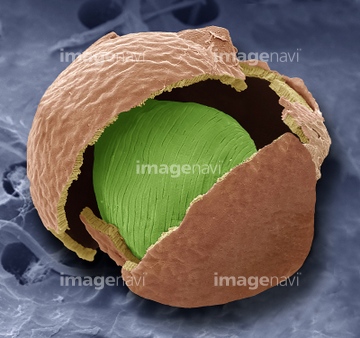
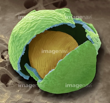
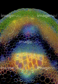
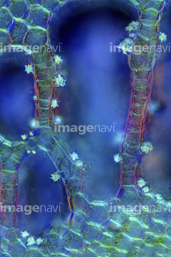
この検索結果には、Christmas rose leaf, SEM、Olive leaf, SEM、Moss cells, light micrograph、Seed pod with seeds (Arabidopsis sp.), SEM、Water milfoil stalk, light micrograph、Water milfoil, light micrographなどが含まれています。
64212739
64212740
64212741
64092140
64092120
64010742
64010743
64010744
64002863
64092119
64050440
64115535
64002864
64210421
64005640
64005641
64115536
64114567
64114569
64145476
64145477

64145484
64148803
64148821
64233688
64124809
64124812
64124813
64124814
64124821
64124823
64124825
64124828
64124831
64124833
64124836
64124843

17212243
64115596
64004043
64005660
64005662
64005663
64260332
64260354
64063704
64250411
64085021
64126577
64115602
64074659
64119747
64009047
64009048
64009049

64260327

64260357
64115520
64115521
64115594

64009115
64009116
64009118
64056946
64056947
64056948
64056949
64225261
64260028
64260029
64260030
64260031
64151007
64002858
20530801
64002853
64117377
64132124
64132125
64107080
64107085
64107086
64107088
64107092
64219420
64219421
64219422
64219423
64219424
64219425
64219426
64219427
64219428
64219429
64219430
64219431
64219432
64219433
64219434
64219435
64219436
64219437
64219438
64219439
64219440
64219441
64219442
64219443
64219444
64219446
64219447
64219448
64219449
64219450
64219451
64219452
64219453
64219454
64219455
64219456
64219457
64219458
64219459
64219460
64219461
64219462
64219463
64219464
64219465
64219466
64219467
64219468
64219469
64219471
64219472
64219473
64219961
64219962
64219965
64219966
64219967
64219968
64219971
64219974
64219975
64219976
64219977
64219978
64219979
64219980
64219981
64219982
64219983
64219986
64219987
64219989
64219990
| 次ページ |
