HOME > 写真 > 科学・テクノロジー > 科学 > DNA・細胞
10,000件の写真素材が検索されました。
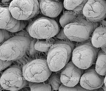
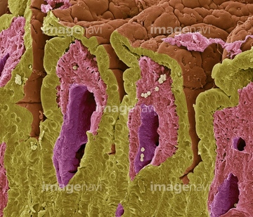
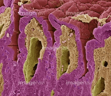
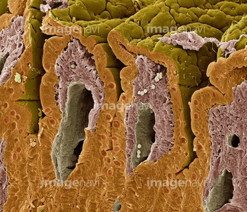
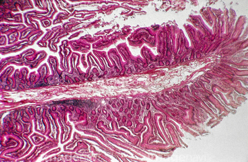
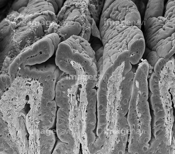
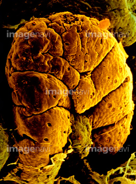
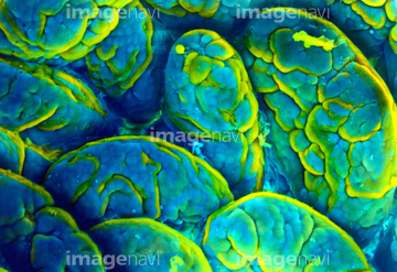
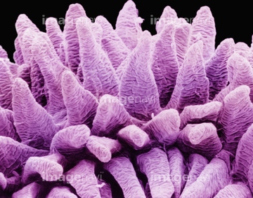
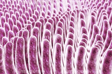
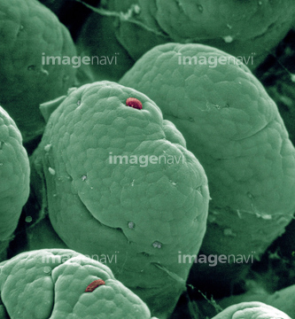
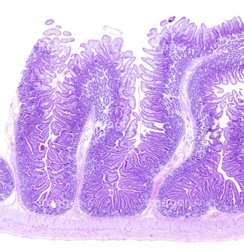
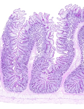
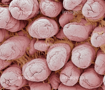
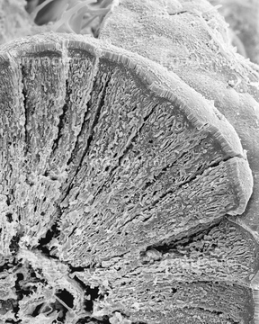
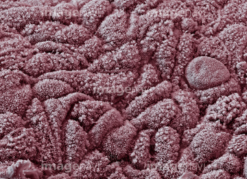
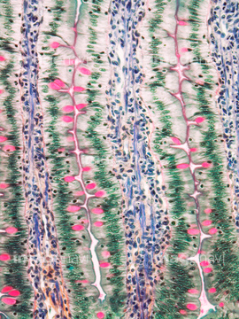
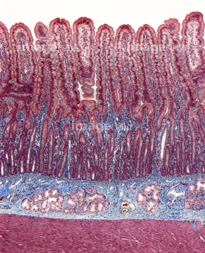
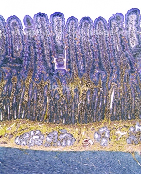
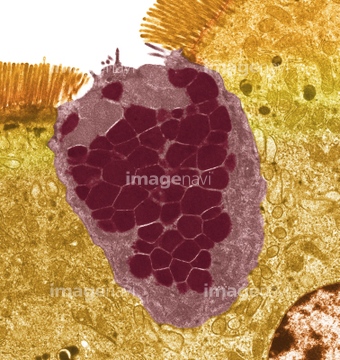
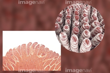
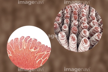
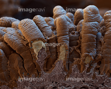
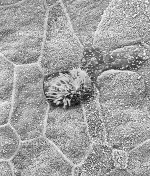
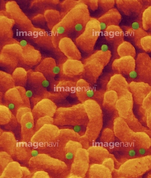
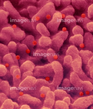
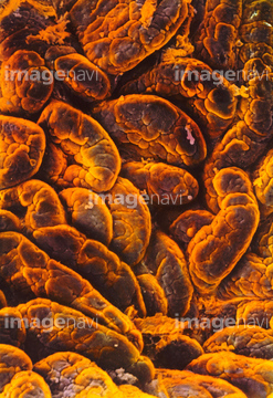
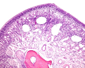
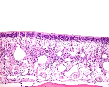
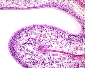
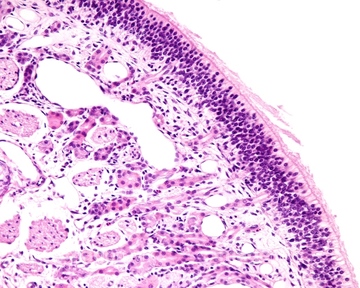
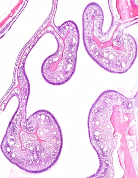
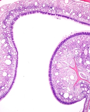
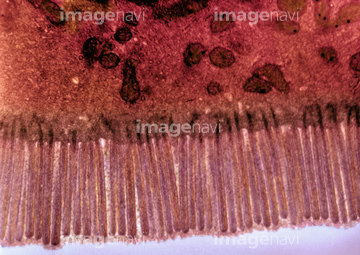
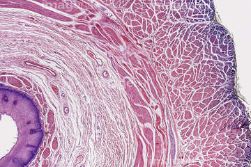
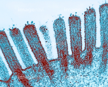
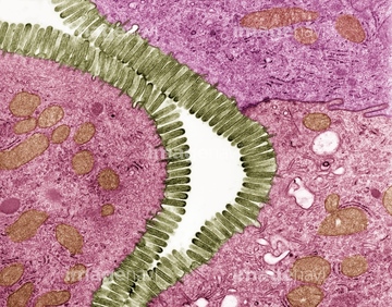
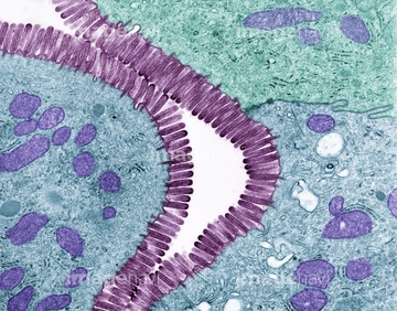
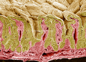
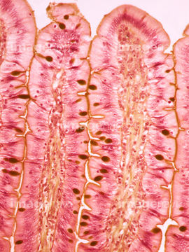
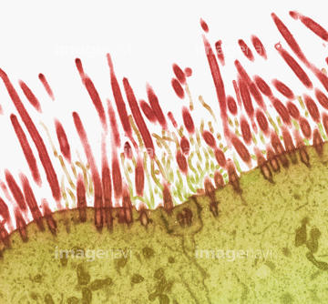
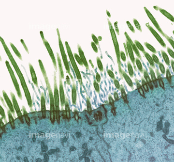
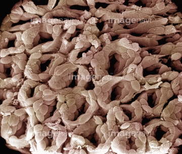
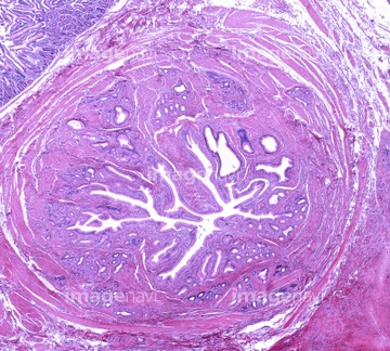
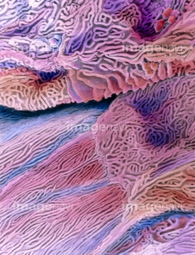
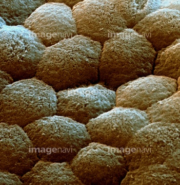
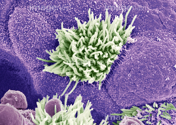
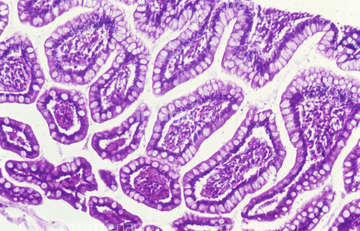
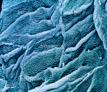
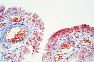
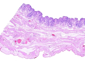
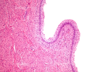
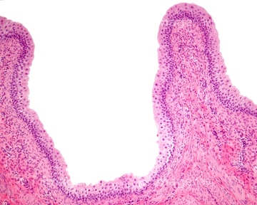
この検索結果には、Intestinal villi, illustration、Intestinal villi cell loss, SEM、Jejunum, light micrograph、Tongue-shaped villi of the jejunum、Intestinal villi, SEM、Small intestine villus and microvilli, SEMなどが含まれています。
64152547
64192859
64192866
64192886
64088314
64152574
64088319
64088323
64262811
64262812
64262814
64262815
64088315

64225140
17202471
17202472
17201424
20523344
20523360
64198564
64234509
64234510
20523355
20523366
64234476
64234477
64234478
64234479
64234508
64225256
17255816
64049910
64196734
64206819

64225133
64075661
64090434
64234667
64234718

64234840
64234841
64234843
17235956
64205888
64205892
64200641
64200647
64061536
64116645
64225131
64176856
64176857
64176858
64049911

64225083
64150338
64132948
64132949
64055781
64209800
17251192
17251193
17251194
64177447
64150091
64021534
64150343
64088321
64088322
64021544
64213913
64213914
64213915
17235955
17200012
17200013
64143715
64143816
64147742
64088343
64055763
64008744
64008745
64008746
64008751
64251226
64190892
64190893
64190894
64190895
64235008
64235009
64235010
64021545
64209773
64209785
64209786
64211723
64211724
64211725
20528355
20528365
20528369
64123467
64123508
64021539
64021540
64021541
64021571
64093980
17202475
17202476
20536138
20536141
64225092
64196715
64136447
64021536
64256459
64234280
64203428
64203450
64203456
64143817
64143818
64143819
64143820
64143821

64133039
64170394
64021533
17200010
17200011
64094080
64197456
| 次ページ |
