HOME > 写真 > 科学・テクノロジー > 科学 > DNA・細胞
10,000件の写真素材が検索されました。
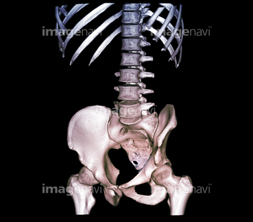
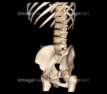
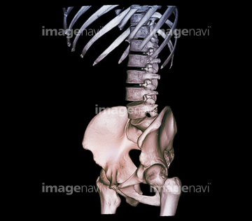
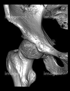
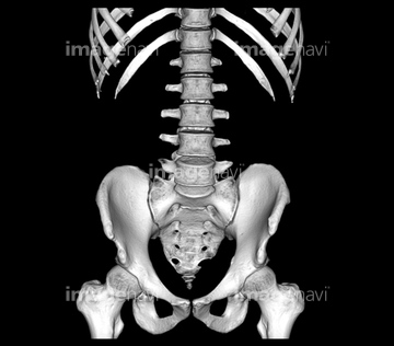
この検索結果には、Human endometrium in secretory phase, light microg…、Healthy lower spine, CT scan、Healthy lower spine and hips, CT scan、Healthy hip joint, CT scan、Sclera, light micrograph、Benign prostatic hypertrophy.などが含まれています。
64154883
64154878
64154881
64154879
64154882
64246907
64078963
64078964
64059784
64154868
64063350
64256471
64246906
64246920
64235141
64235142
64059783
64159966
64065774
64256348
64256355
64154889
53157300
53157301
53157302
53157303
64246120
64246121
64246176
64198948
64198960
64198961
64198962
53157252
53157253
53157254
53157255
53157349
53157350
53157351
53157352
64200575
64170393
64116654
64116675

64162976
64162977
64180134
64180236
64180237
64184847

64225308
64159057
64222730
64184337
64256497
64119245
64243665
64256396
17256236
64112139
64234962
20530725
64151858
64151859
64151860
64151861
64154872
64154873
64065583
64065983
64065987
64116648
64065589
64065615
64062937
64062938
64062969
64062970
64062971
64062972
64062973
64062974
64062975
64062976
64062977
64062982
64246118
64170387
64170388
64170389
64170390
64170395
64170396
64170397

64246490
64246491
64246492
64057702
64264227
64264228
64176635
64194973
64194974
64219696
64219697
64219698
64219699
64219709
64219710
64219711
64257169
64257170
64257171
64257172
64197111
64008670

64180354
64216421
20542509
64159097
64159119
64159120
64159121
64225384
64213834
64213835
64151856
53157248
53157249
53157250
53157251
64184329
64184338
64152577
64150085
64065616

20562628
64048824
64065591
64065614
64065618
64065619
64065623
64065624
64065775
64065784
64065984
64236962
64236963
| 次ページ |
