HOME > ژتگ^ > ژY‹ئپEٹآ‹«–â‘è > ƒTپ[ƒrƒX‹ئ > ˆم—أپE•ںژƒ‹ئ
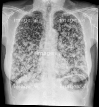
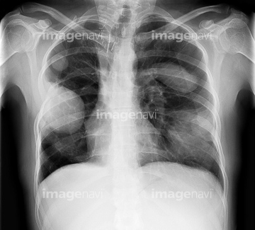
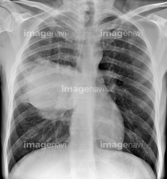
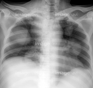
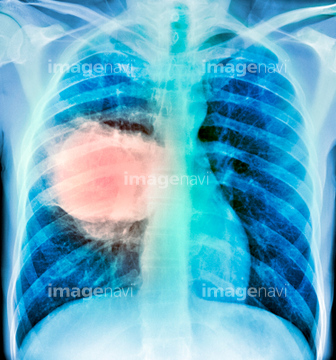
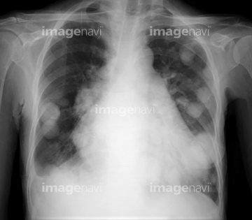
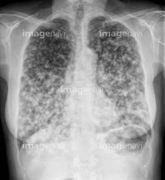
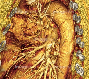
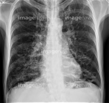
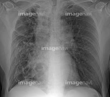
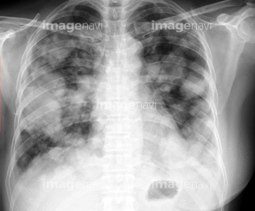
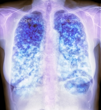
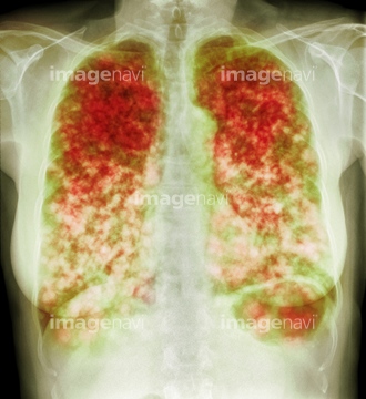
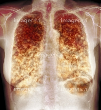
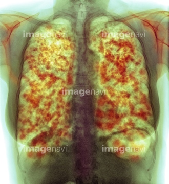
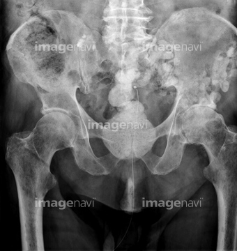
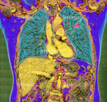
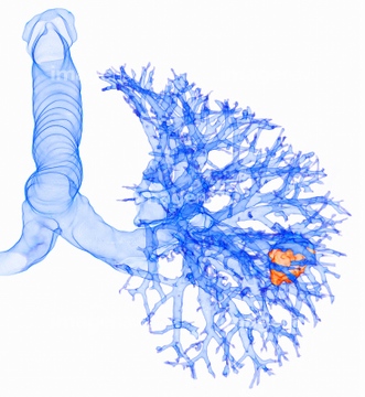
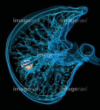
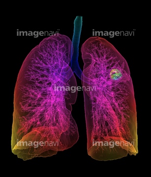
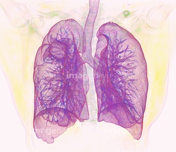
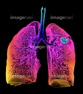
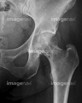
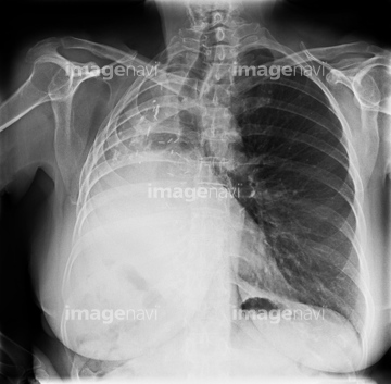
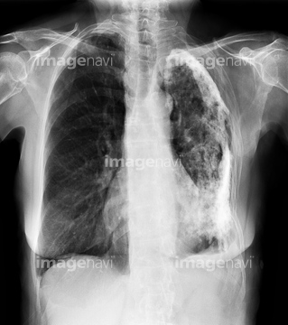
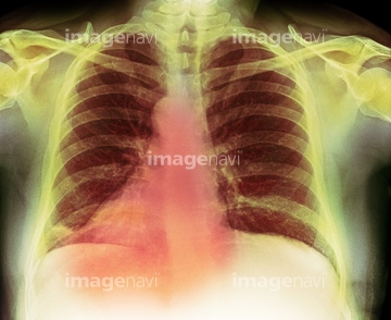
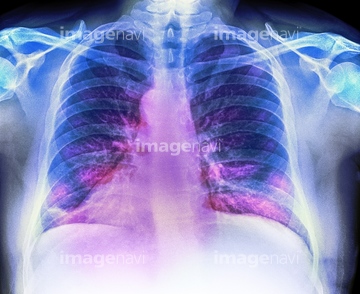
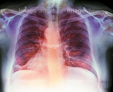
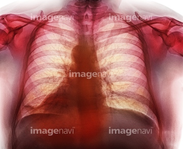
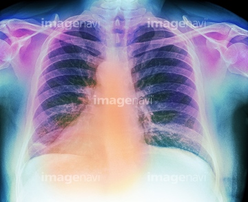
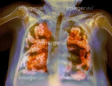
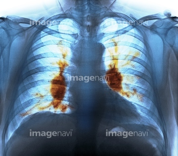
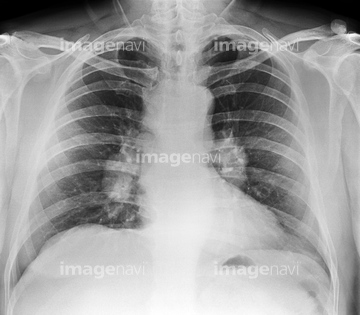
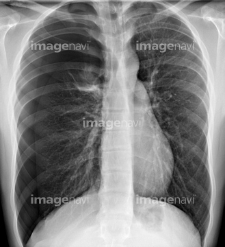
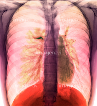
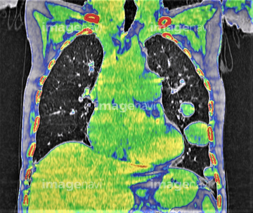
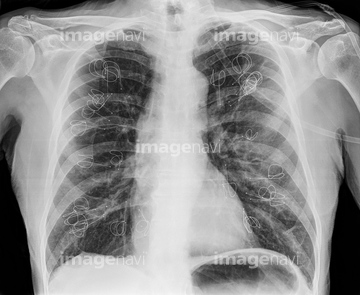
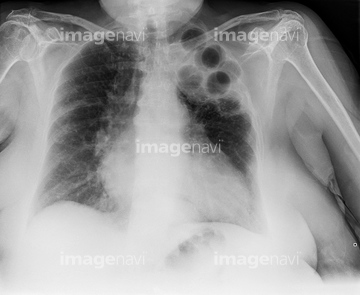
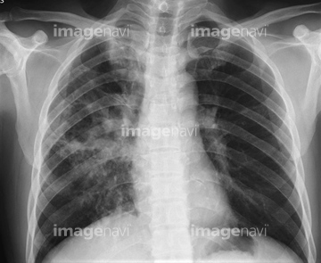
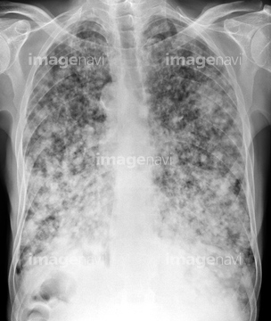
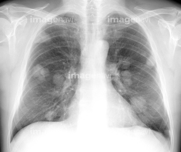
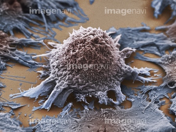
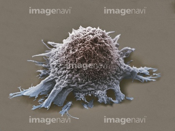
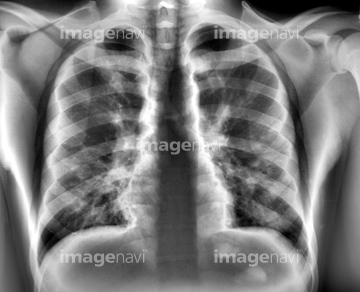
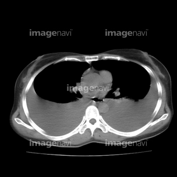
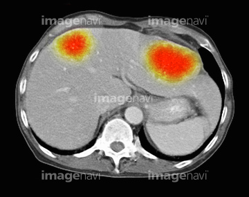
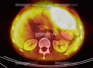
10,000Œڈ‚جژتگ^‘fچق‚ھŒںچُ‚³‚ê‚ـ‚µ‚½پB
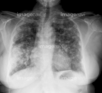
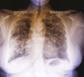
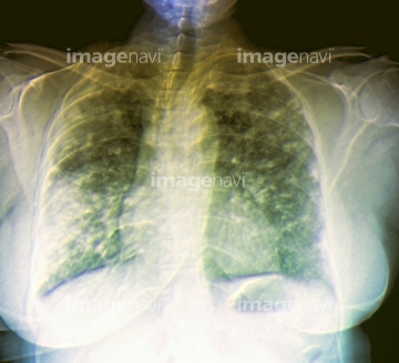
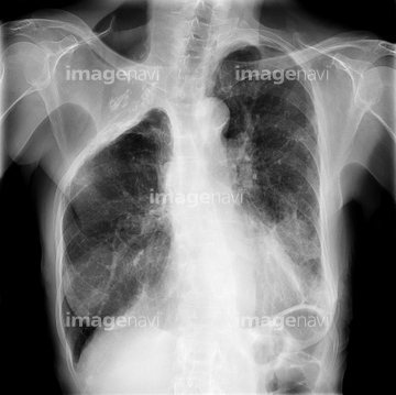
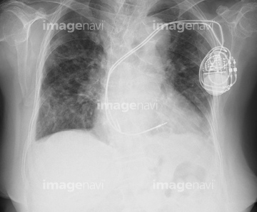
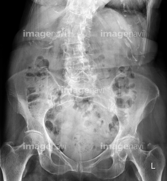
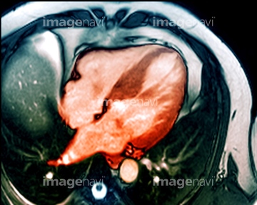
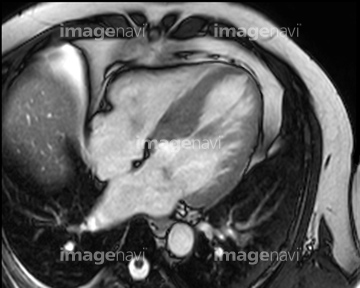
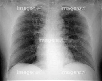
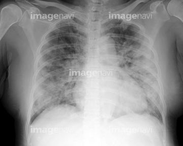
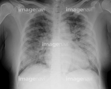
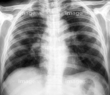
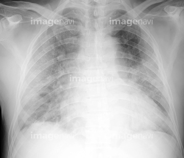
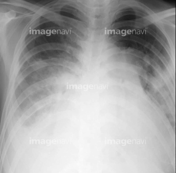
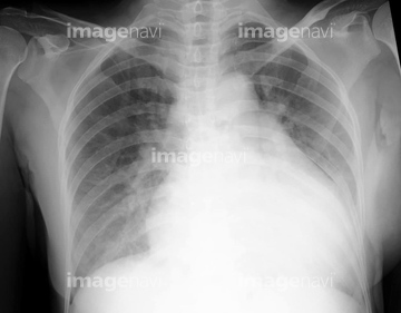
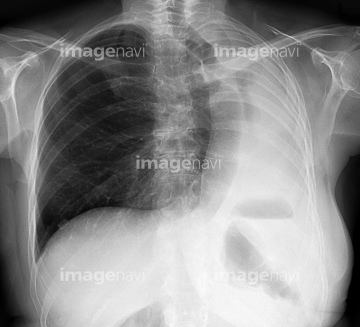
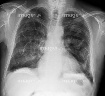
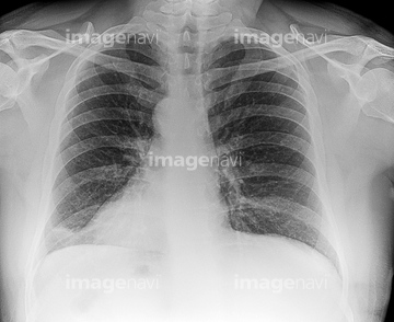
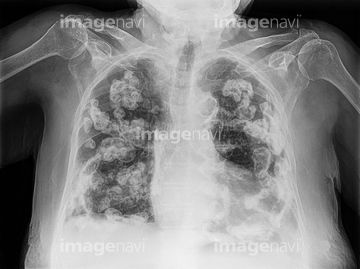
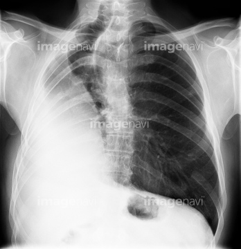
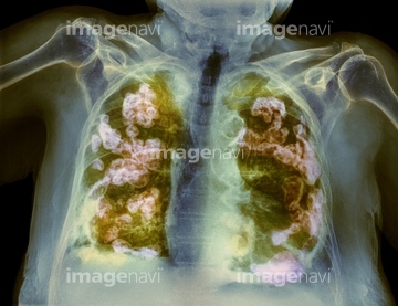
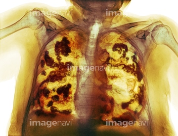
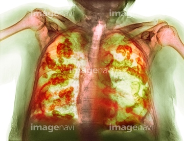
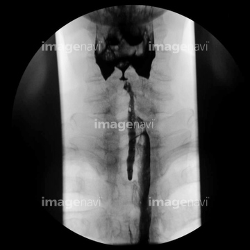
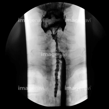
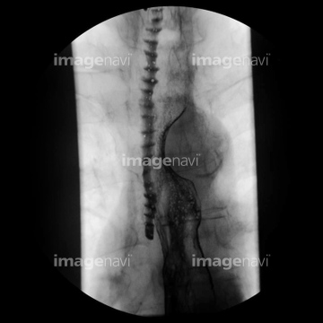
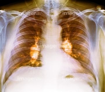
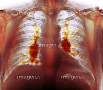
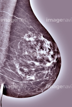
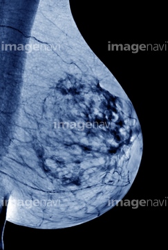
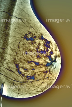
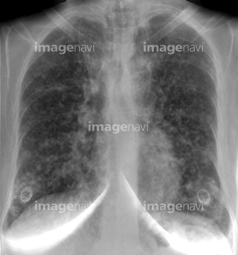
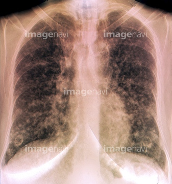
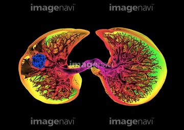
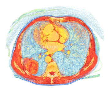
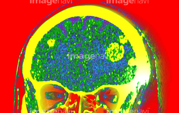
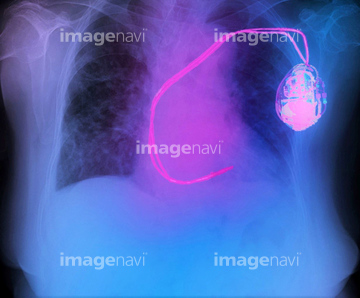
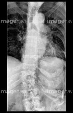
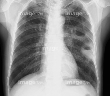
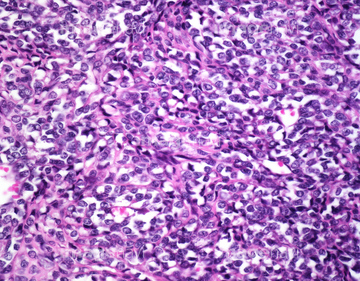
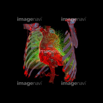
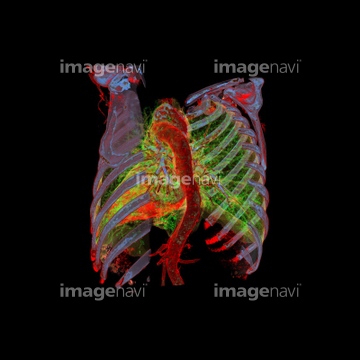
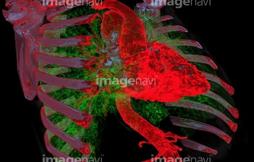
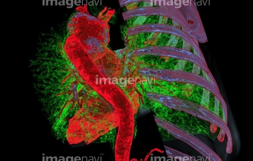
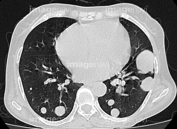
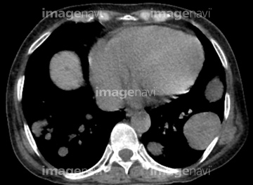
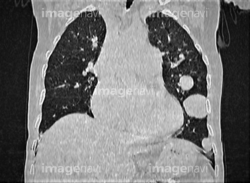
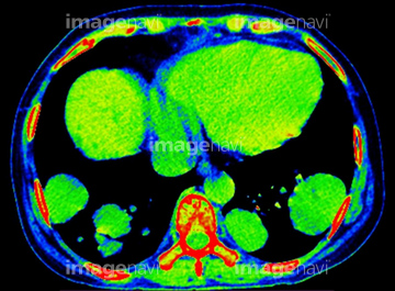
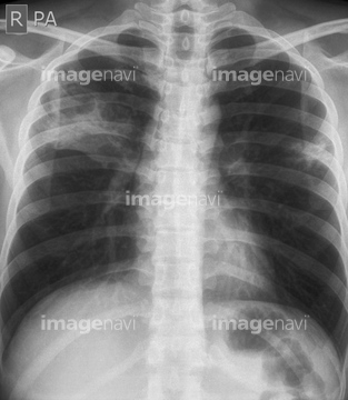
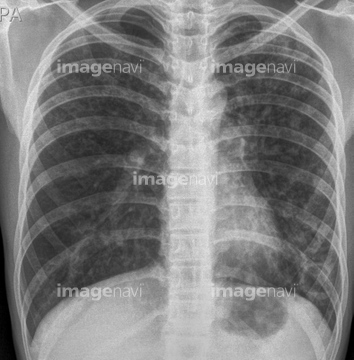
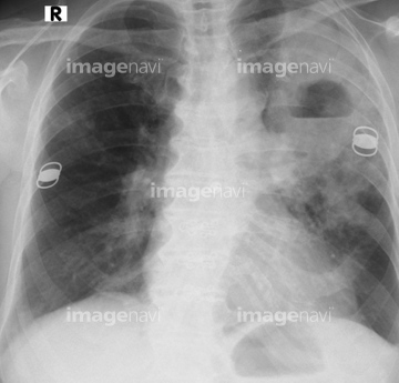
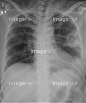
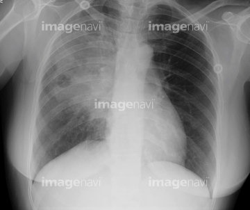
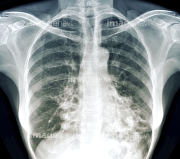
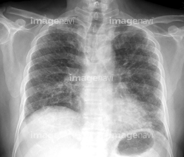
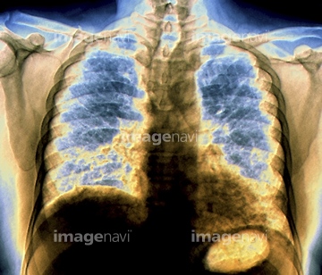
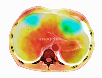
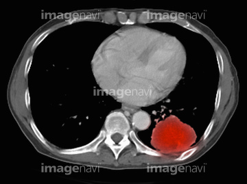
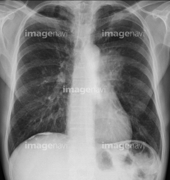
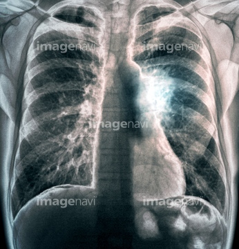
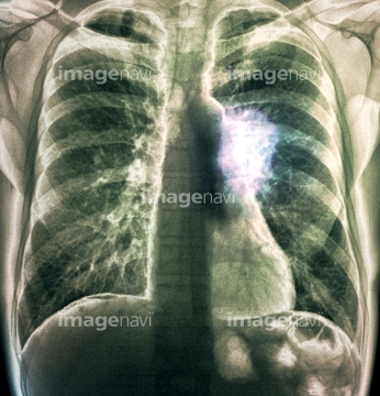
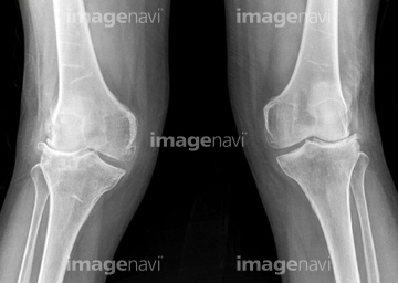
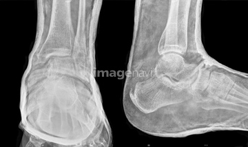
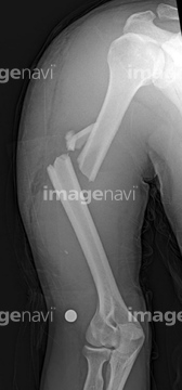
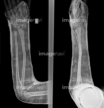
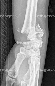
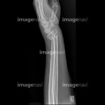
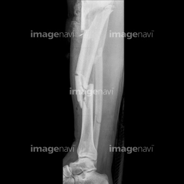
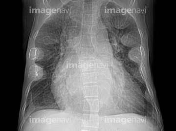
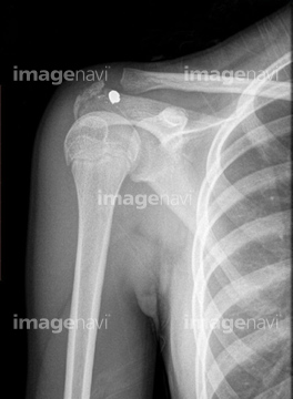
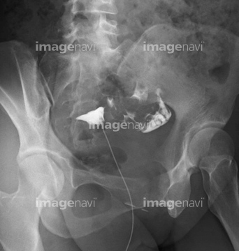
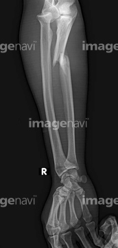
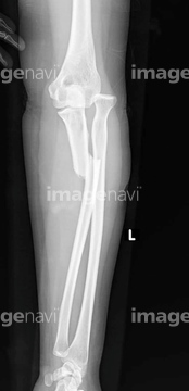
‚±‚جŒںچُŒ‹‰ت‚ة‚حپAEnlarged heart and pulmonary oedema, X-rayپALeft pneumonectomy, X-rayپAPleural thickening due to asbestos, X-rayپAReversed heart position, X-rayپABilateral pleural calcification, X-rayپARight pneumonectomy, X-ray‚ب‚ا‚ھٹـ‚ـ‚ê‚ؤ‚¢‚ـ‚·پB
64162668
64159403
64087127
20543726
64165243
64165244
64165245
64159397
64087125
64166777
64153244
64153245
64174834
64174835
64174836
64174837
20582123
20582179
20543727
64166773
64166774
64166775
64166776
64153254
64174832
64174833
64159426
64159427
64159428
64159429
64159430
64159431
64149471
64159903
64159904

64159905
64153234
64153235
64153236
20582155
64159411
20543681
20543683
20543684
20543685
20543686
64159389
64159399
64159417
64159423
64159439
64160596
64160597
64160598
64160599
64160600
64164388
64164389
64164390

64164391
64164392
64087151
64087153
20543692
20543721
64219590
64220130
64220131
64220132
64137965
64137966
64153237
64153255
64250744
64250745
64250746
64155037
20582156
64162682
20582135
64260885
64260886
64260887
64260888
64260889
64260890
64168545
64168546
64188544
64188545
64188546
64188547
64188548
64188549
64188550
64188551
64188552
64194419
64194420
64192653
20562180
20562181
20562385
20562387
20562388
20562389
64151753
64165507
64165508
64064320
64064329
64174034
64174048

64176152

64176153

64176154

64176155

64176156
64176157
64179825

64179826
64179827

64179828
64261243
64261244
20564326

64221196
64221197
64221198
20543696
20582122
20568284
20548371
20548372
20548373
20548374
20548375
20548376
20548381
20548382
20548383
20548388

20548389
20548392
20548394
20548395
20548396
20548397
20548398
20548400
20548408
20548409
20548413
| ژںƒyپ[ƒW |
