HOME > 写真 > 人物 > 外国人 > 男性
10,000件の写真素材が検索されました。
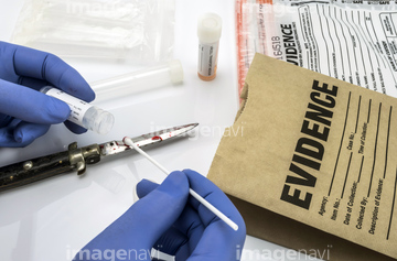
この検索結果には、Brain specimen preparation、Fractured lumbar vertebra, CT scan、Crime scene、Fractured ankle bone, CT scan、Dislocated lens of the eye, CT scan、Lawnmower hand injury, X-rayなどが含まれています。
64163244
64017999
64145829
64017998
64017997
64145819
20574339
20574341
64242421
64017991
20569526
20569551
20569561
20569550
20574375
20574380
20574382
20573745
20573746
20555766
20555767
64223389
64017995

64159430
64017992
64151245
64017975
64017984
64017985
64018006
64018014
20574340
20574376
20574383
20574384
20574385
20574386
64203163
64203167
20573742
20573743
20573744
64145845
64197236
64197237
64018013
20512554
20512555
20512556
20564323
64159398
64159400
64159401
64159407
64159432
64162669
64162683
64145826
64145830
64205375
64205376
64204800
64162691
64224125
20567505
64165602
64165603
20546202

20546203
20546204

20563371
20574314

20574337
20574361

20574372

20574400
17280190
17280191
64018010
64018011
64018012
64145859
64145863
64145875
64245962
52218624
16932631
64151573
20566621
20568287
20573748
64159269
64159273
64159275
64159276
64177844
64177845
64177846
64177847
64177848
20573161
64203164
64151883
64151884
64148049
64208777
64208778
64208779
20574381
20564382
20566744
20566745
20566746
64160610
64160612
64159270
64159271
64159272
64159274
64159277
64159278

64188222
64205332
64205333
64162670
64205330
64205331
64205339
64205340
64205341
64205342
64205343
64205344
64205345
64205346
64205347

64205348
64205349
64205350
64205351
64205353
64205354
64205355
64205356
64205358
64205359
64205360
64205361
64205362
64205369
64205370
64205371
64205372
64205373
64205374
64205378
64205379
64205380
64151244
19997132
20512557
20512558
64204799
20548371
20548373
20548375
| 次ページ |
