HOME > 写真 > 科学・テクノロジー > 科学 > DNA・細胞
10,000件の写真素材が検索されました。
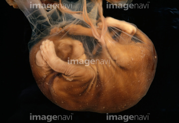
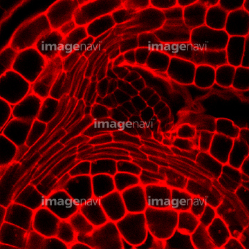
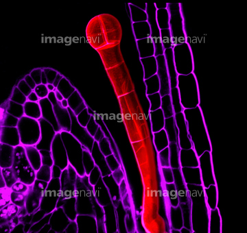
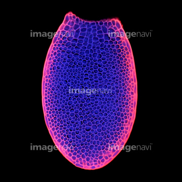
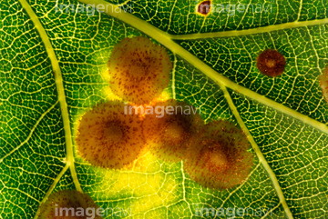
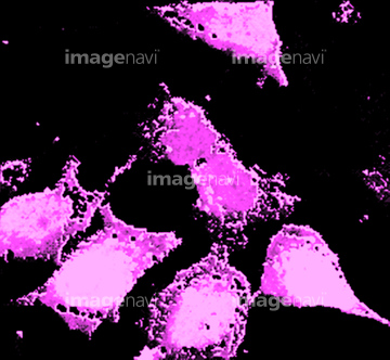
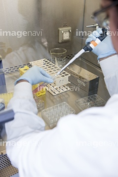
この検索結果には、Monkey foetus、Plant embryo, light micrograph、Arabidopsis thaliana embryo, micrograph、Common spangle galls on an oak leaf、Neuroblast cells, fluorescent micrograph、Stem cell embryology researchなどが含まれています。
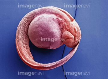
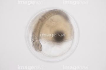
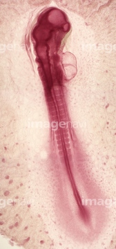
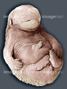
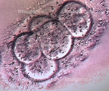
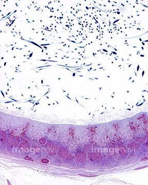
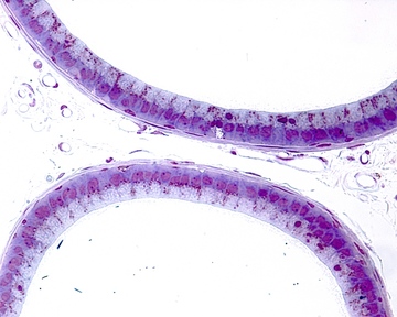
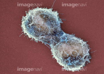
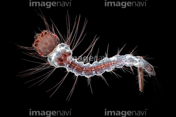
64261170
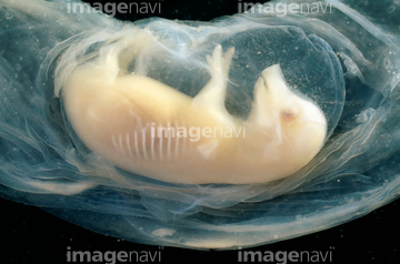
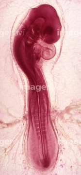
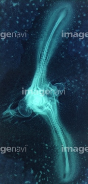
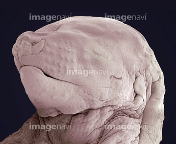
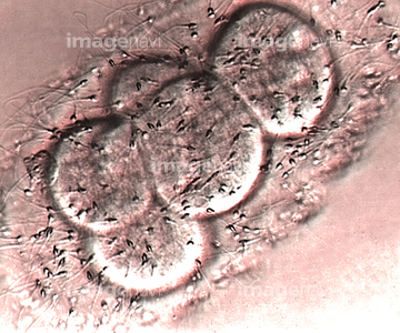
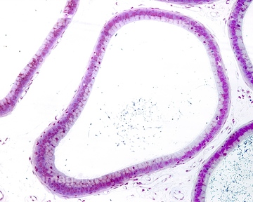
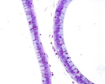
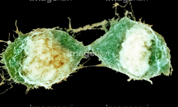
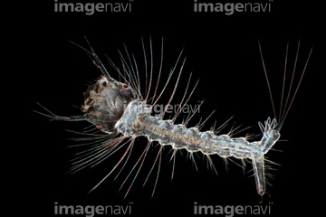
64261171
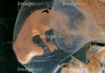
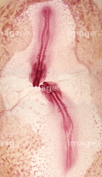
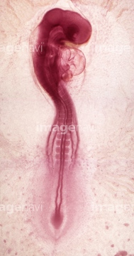
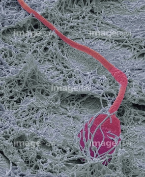
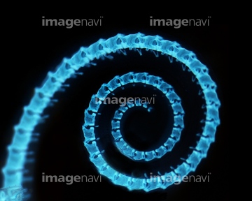
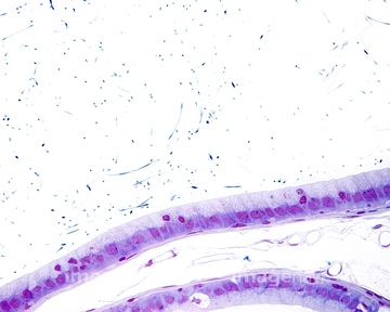
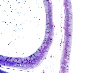
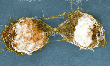
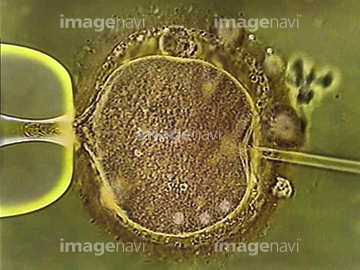
64261172
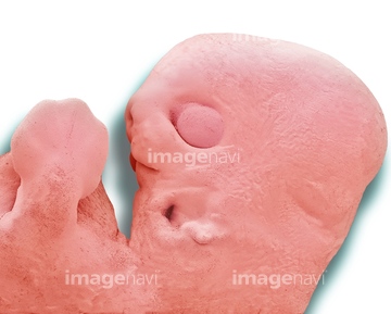
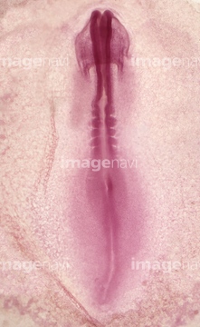
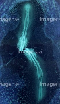
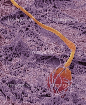
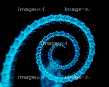
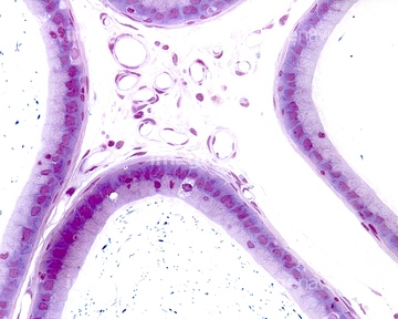
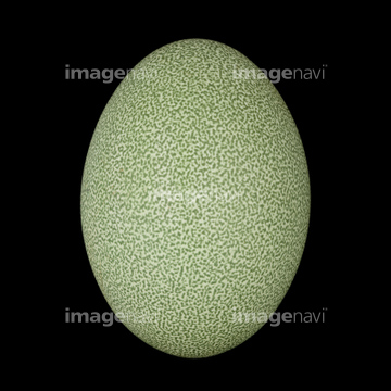
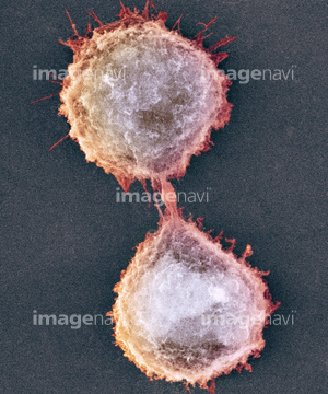
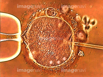
64261173
64089201
64055598
64119231
64089205
64152751
64152752
64022139
64089199
64022133
64225095
64022128
64022131

64108882
64108908
64152749
64152754
64152757
64044638
64044639
64002909
64003101
20532073
20532081
64022140
64055599
64055600
64163530
64119230
64085311
64121354
64022129
64022130
64022132
64010998
64010999
64003715
64152755
64152758
64058235
64244183
64244186
64244202
64109513
64109516
64109523
64109550
64263893
64085230
64034997
64116829
64060353
64051867
64253139
64150301
64003076
64003543
64003546
64040557
64040559
64040881
64098167

64117275
64263894
64003250
64152740
64152747
64152748
64152750
64152753
64152756
64196737
64032663
64180225
64124387
64124388

64225140
64225256
22432875
64226868
64191458
64083855
64083856
64129601
64129603
64129604
64129608
64129614
64129615
64129617
64063606
64063607
64049707
64008754
64008755
64008756
64132036
64132049
64105451
64105457
64105461
64105472
64105480
64106165
64245992
64245993
64245994
64124570
64119226
64176756
64176757
64176758

64010761
64010762
64010764
64010765
64010767
64010768
64010770
64010771
64045959
64074952
64075097
64075143
64075451
64037750
64167028
64167034

64167037
64180318
64180319
64180327
64180329
64180334
64180337
64109526
64109551
64085316
64085323
64150016
17238432
64099928
64156134
64156136
| 次ページ |
