HOME > 写真 > 医療・福祉 > 医療 > 病院・クリニック
10,000件の写真素材が検索されました。
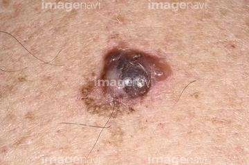
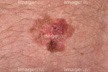
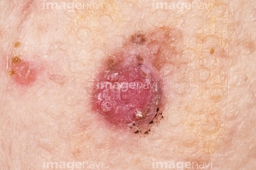
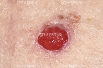
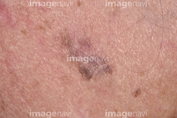
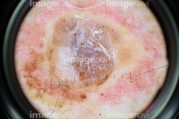
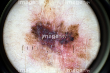
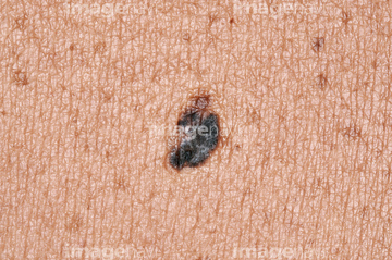
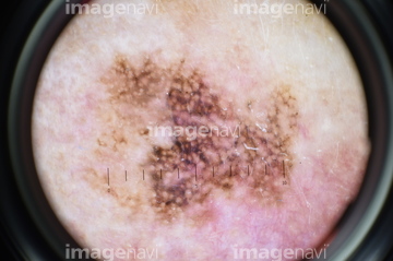
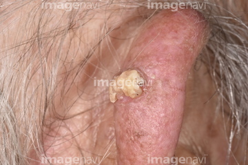
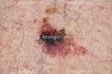
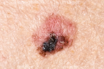
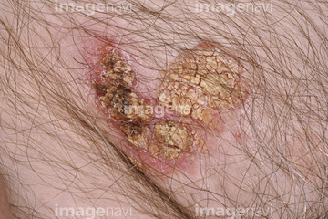
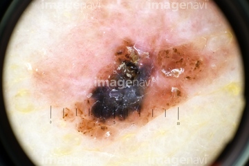
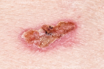
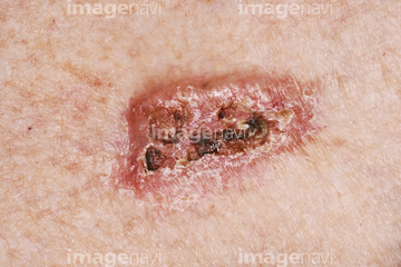
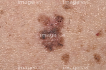
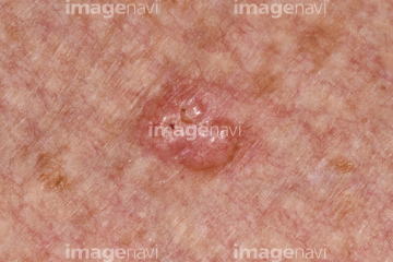
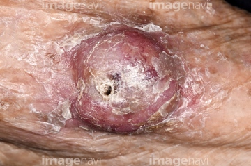
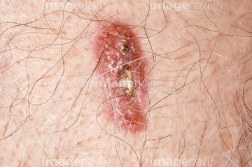
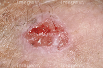
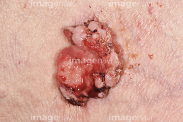
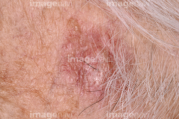
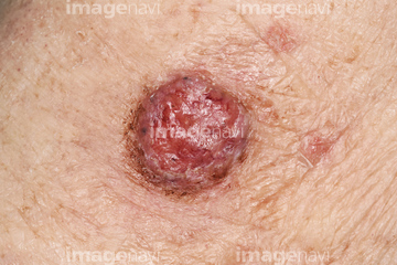
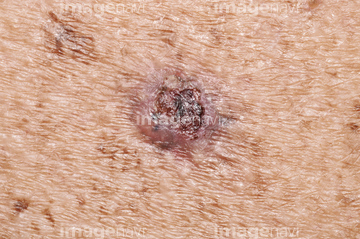
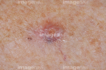
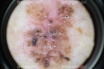
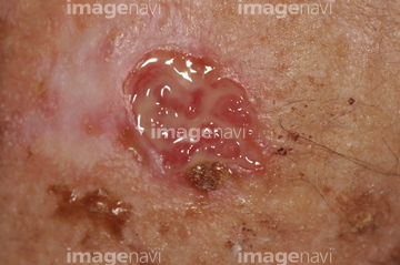
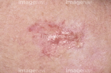
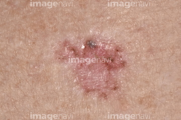
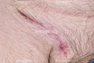
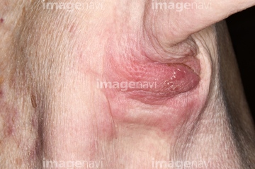
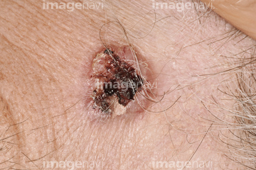
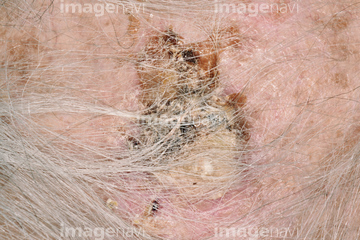
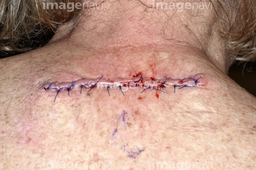
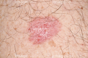
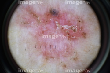
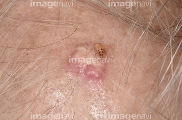
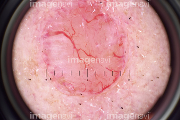
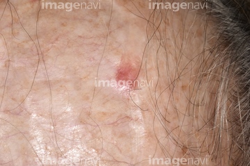
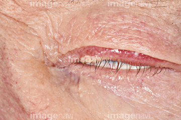
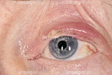
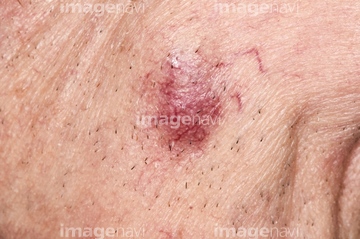
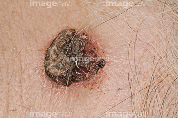
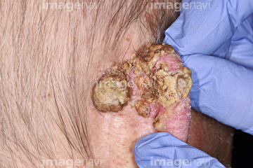
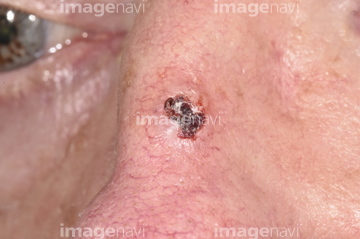
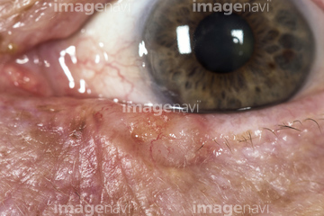
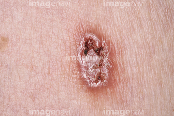
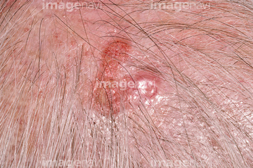
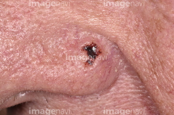
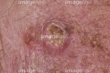
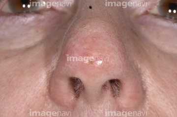
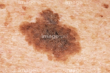
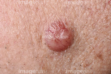
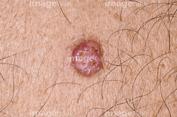
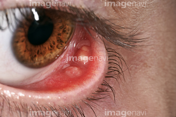
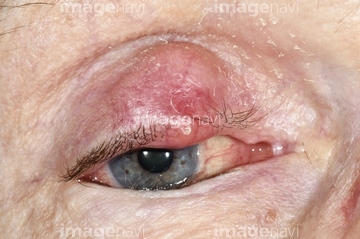
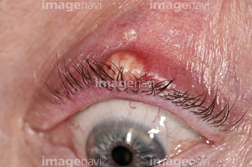
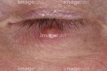
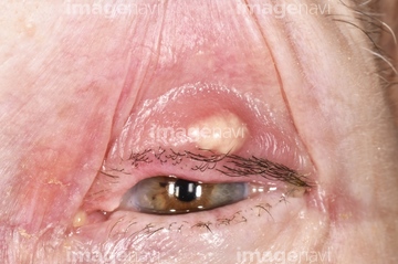
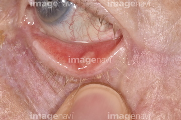
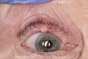
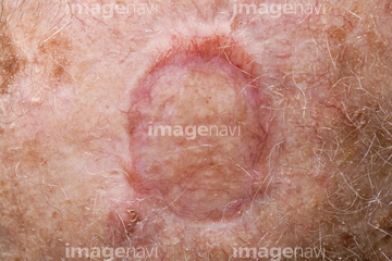
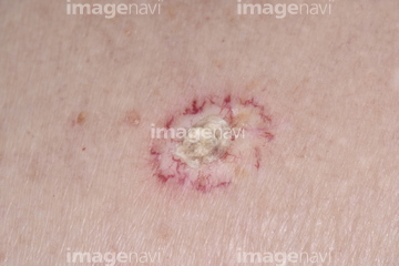
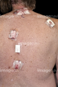
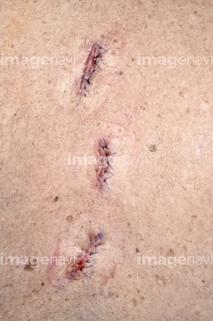
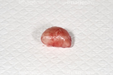
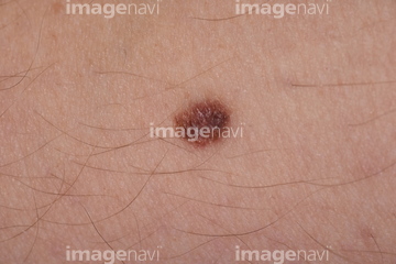
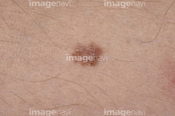
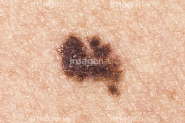
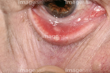
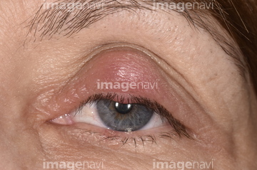
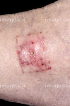
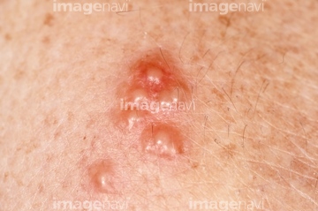
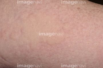
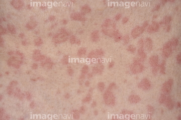
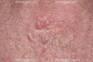
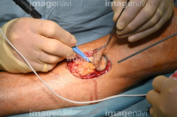
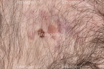
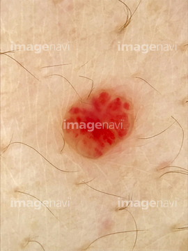
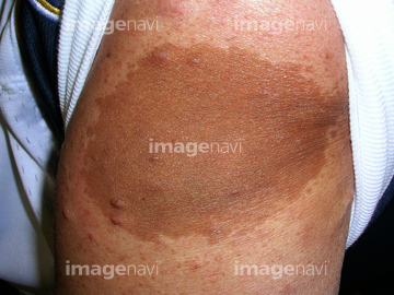
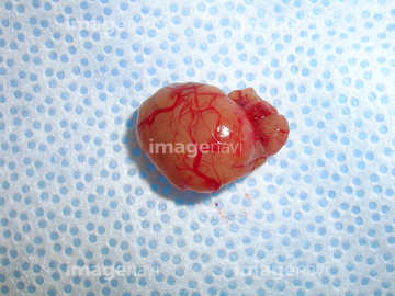
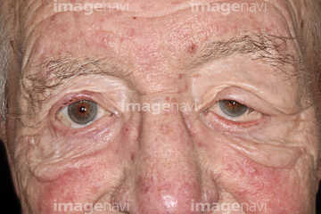
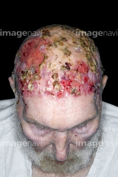
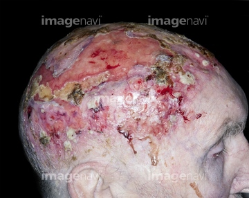
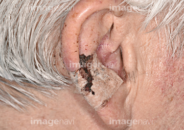
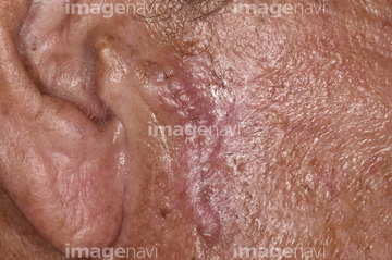
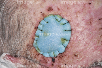
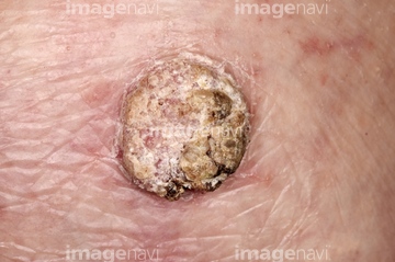
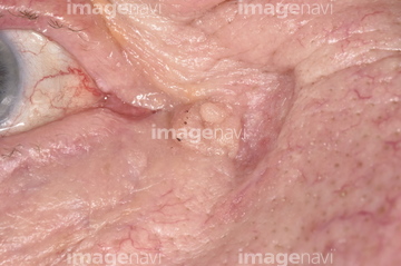
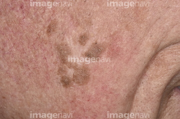
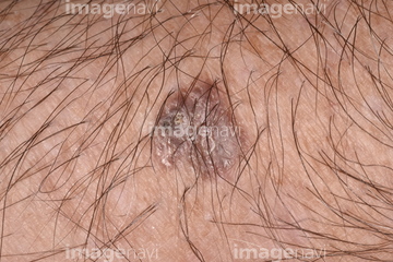
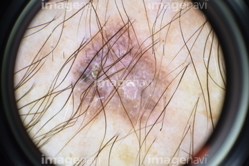
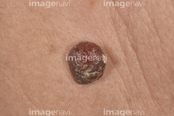
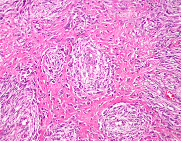
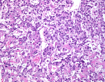
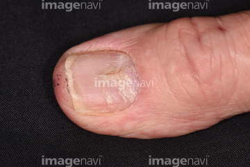
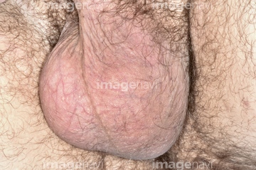
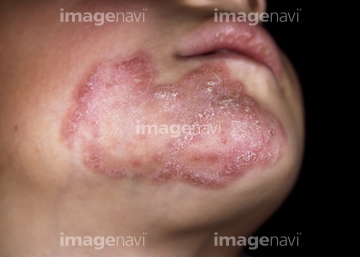
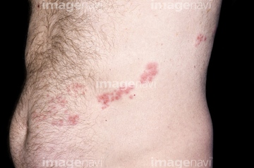
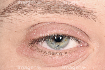
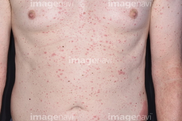
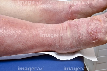
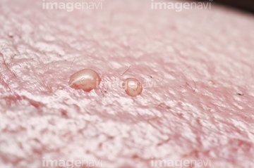
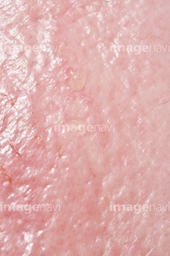
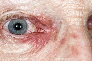
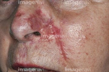
この検索結果には、Chalazion abscess on an eyelid、Merkel cell carcinoma、Squamous and basal cell carcinomas before treatmen…、Squamous cell carcinoma、Basal cell carcinoma、Ulcerated basal cell carcinomaなどが含まれています。
64168051
64168050
64168045
64168124
64190946
64188283
64158708
64158709
64188286

64188287
64168049
64168044
64188288
64168048
64158710
64159312
64159313
64159314
64159315
64158711
64158702
64168128
64188284
64188293
64158700
64158701
64168126
64168127
64168135
64168137
64168138
64168139
64188285
64188289
64188290
64188291
64168043
64158712
64168140
64190968
64168125
64188292
64188295
64168047
64158623
64168052
64168053
64158645
64168141
64191320

64158703
64159248
64168122
64168136
64188282
64188294
64188296
64158617
64168046
64168143
64168151
64190950
64168152
64188379
64158715
64188307
64188309
64190969
64190970
64158637
64253410
64253412
64225824
64168145
64188377
64253411
64253414
64225825
64153214
64137912
64149471
64159397
64159403
64158707
64225842
64225843
64158635
64159426
64159428
64159429
64159431
64153244
64153245
64159905
64207288
64253256
64152373
64152374
64253257
64188437
64158606
64158607
64159059
64188211
64158566
64158608
64159061
64188208
64188217
64204923
64252696
64252689
64252732
64167969
64167970
64159427
64159430
64158634
64188227
64188228
64188240
64188241
64188278
64153213
64207294
64244388
64190948
64146833
64159056
64168154
64188210
64244453
64151226
64158609
64158618
64113184
64113185
64260885
64260886
64260887
64260888
64260889
64260890
64145817
64145818
64190951
64168144
64253413
64257334
64257338
64260600
64260601
64260602
64257279
64254472
| 次ページ |
