HOME > 写真 > 医療・福祉 > 医療 > 手術
10,000件の写真素材が検索されました。
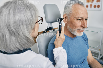
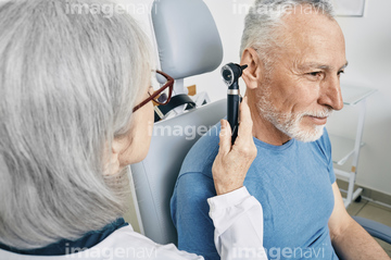
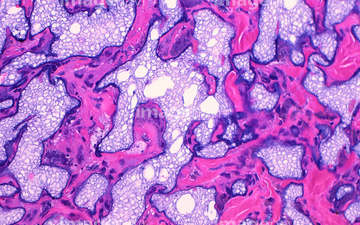
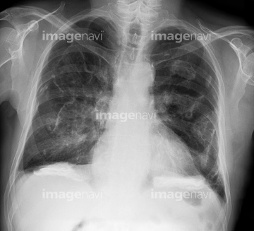
この検索結果には、Acne rosacea、Ovarian cyst, endoscope view、Aplasia cutis congenita、Dysplastic naevus, dermascope image、otolaryngologist examines the ear of the patient、Left pneumonectomy, X-rayなどが含まれています。
64168064
64168063
64153408
64153409
64161934
20567714
20567715
20567716
20567717
20567718
20559858
20559859
20562631
64159417
20566126
20566127
20547611
20547612
20547615
64153411
20567721
20567724
20567725
64153393
64159423

64008486
64144855
64144856
64144857
64148446
64148447
20560269
20560273
64260567
64159421
20549083
20549084
64153390
64153391

20566118
20566128
20566129
64177842
64177843
64154872
64154873
64153410
64212576

64159389

20566239

20566240

20567761
17203029
17203030
17203097


64256357
64153412
64225753
64168152
20560351
64153392
64177859
64177860
64177861
64188199
64158637
20562926
20562927
20543681
64159399
64153400
64153401
64162696
64162697
64260586
64260588
64260589
64260590
64260591
17203473
17203582
64021022
20562937
20547581
20547582
20547583
20547584
20547585
20549157
20566117
20547599
20549202
20567730
64188379
64021020
64021021
20559943
64159432
20566125
64019226
64243136
20532075

20532084
20582159
20582160
17277205
64153394
64153395
64153396
64153403
64153404
64153405
64135842
64135843
64135844

64186255
64256329
64256335

64256351
64256352
64256353

64256362
64256367
64256370
64256410
64256438
64256448
64256487
| 次ページ |
