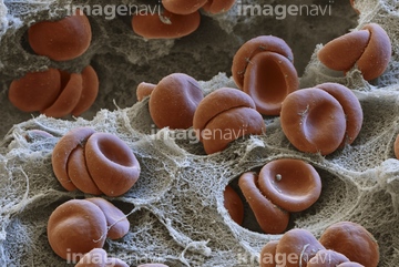
HOME > 写真 > 科学・テクノロジー > 科学 > DNA・細胞
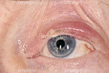
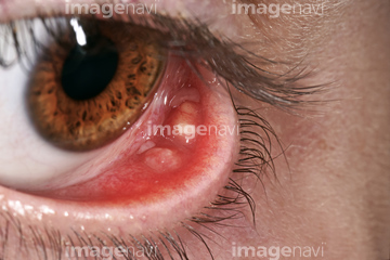
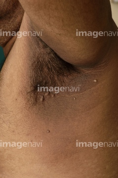
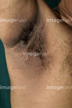
10,000件の写真素材が検索されました。
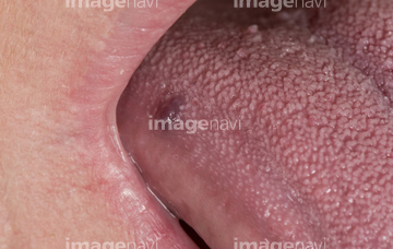
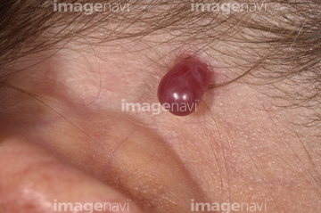
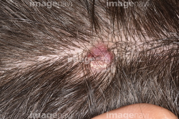
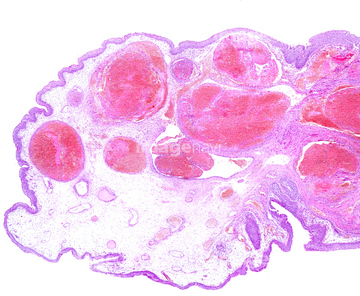
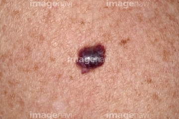
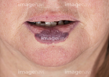
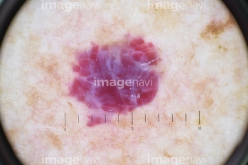
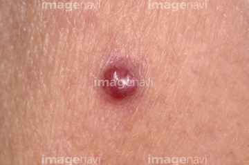
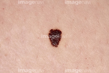
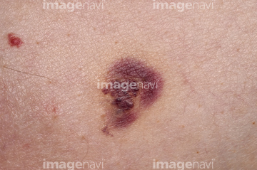
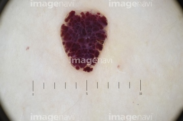
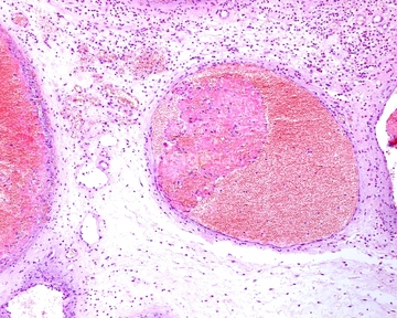
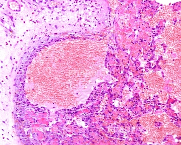
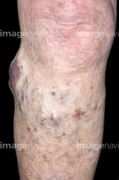
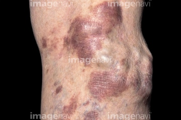
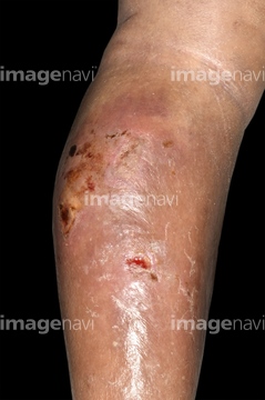
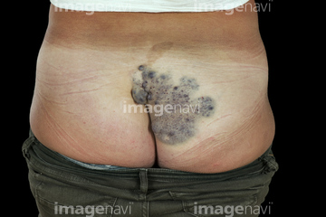
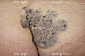
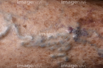
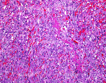
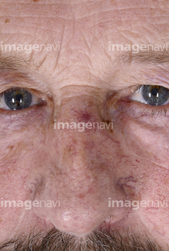
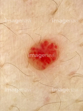
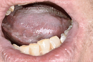
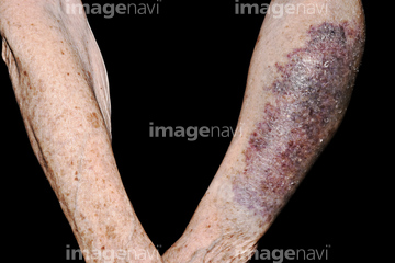
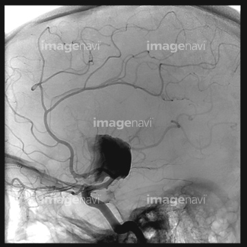
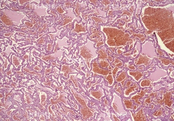
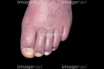
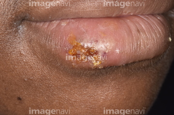
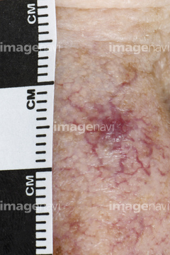
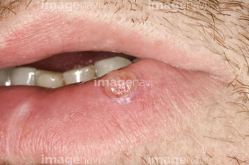
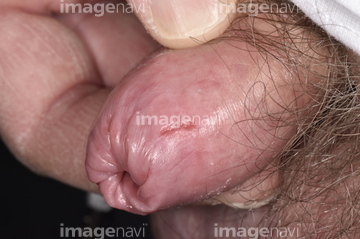
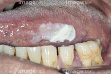
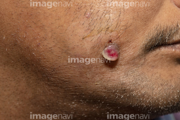
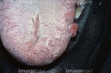
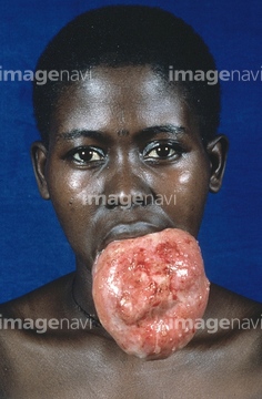
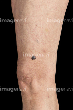
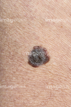
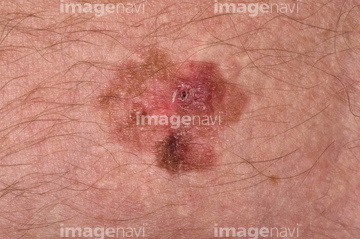
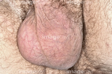
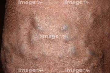
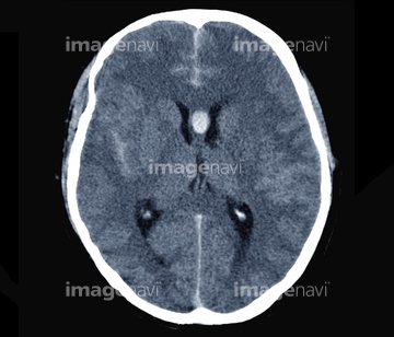
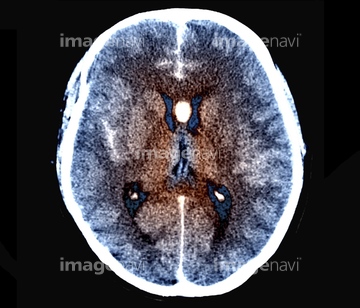
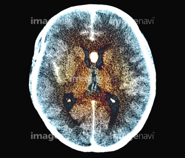
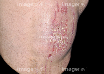
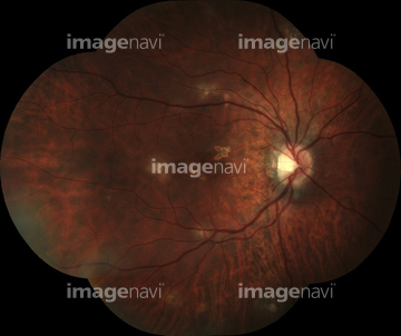
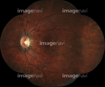
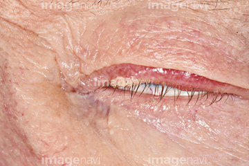
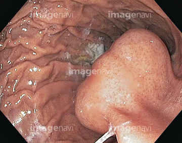
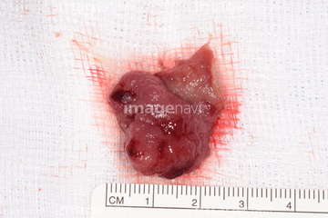
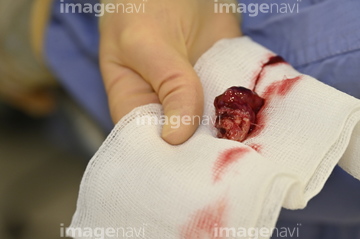
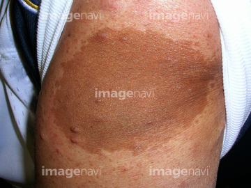
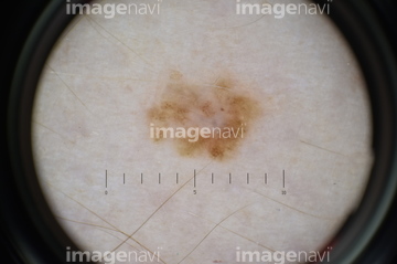
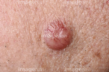
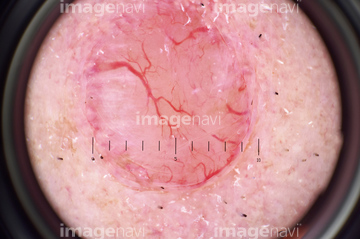
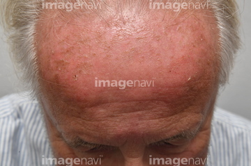
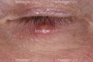
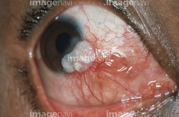
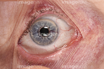
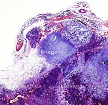
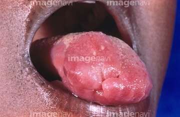
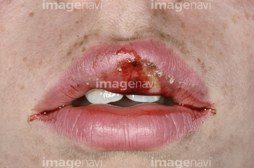
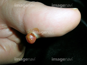
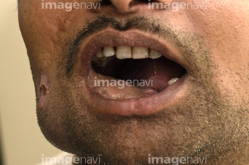
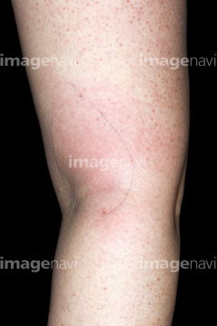
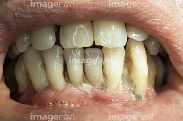
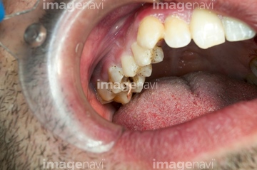
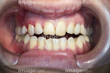
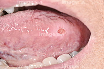
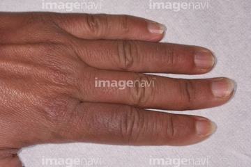
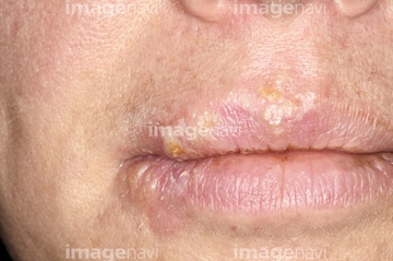
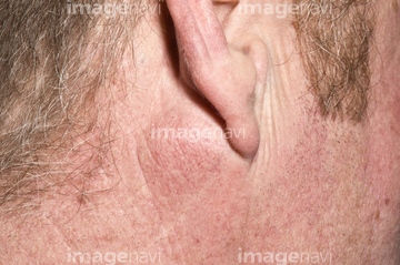
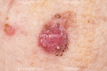
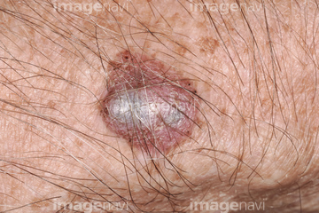
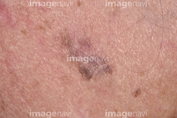
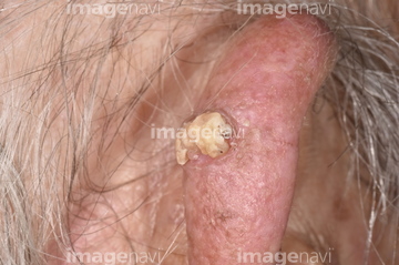
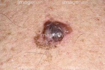
この検索結果には、Cold sore on lip、Angioma、Squamous cell papilloma on a woman's lip、Pyogenic granuloma、Inflamed foreskin in diabetes、Leukoplakia under the tongueなどが含まれています。

64177831
64167391

64159295
64159296
64188267
64188273
64173406
64159303
64173404
64173408
64204876
64173409
64158619
64158620
64159050
64165742
64165743
64188226
64260526
64151582

64252696
64168123
64158627

64159003
64159004
64165744

64165745
64165746
64165497

64192659
64153228
64204877
64158625
64158626
64158628
64158629
64168168
64188410
64188437
64151585

64172381
64188276
64159307
64219667
64264818
64264747
64159312
64159313

64159314
64188283
64190948
64222679
64222698
64152558
64173437
64129796
64149482
64149483
64212393
64212394
64151193
64151194
64151195
64151196
64168144
64252663
64188277
64129820
64205894
64205902
64096091
64174404
64167382
64167383
64168052
64168053
64168151

64152290
64152373
64152374
64259854
64259895
64252689
64252732
64253410
64253411
64253412
64253414
64168046
64168047
64188379
64264999
64168153
64264696
64174399
64253986
64152361
64191181
64263270
64139323
64165161
64168166
64188199
64188265
64143024
64143025
64143026
64143027
64150080

64151198
64192660
64192661
64192662
64188409
64129642
64129643
64140919
64140920
64140921
64152583
64152585
64205330
64205331
64230414
64230415
64137926
64137927
64137928
64188544
64188545
64188547
64188548
64188549
64188550
64188551
64188552
64192653
64192654
64071991
64072066
64123604
64253251

64253252
64159059
64158707
64158708
64158709
64159315
64168050
64168051
64168124
64188286

64188287
64190946
| 次ページ |
