HOME > 写真 > ライフスタイル > 体調
10,000件の写真素材が検索されました。
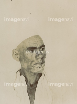
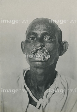
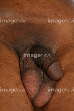
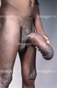
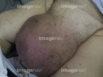
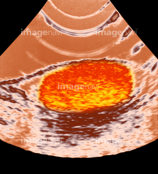
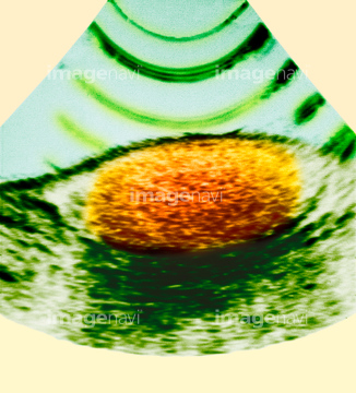
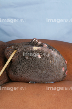
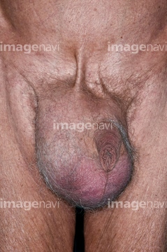
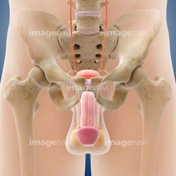
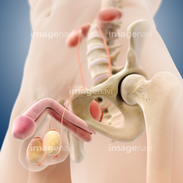
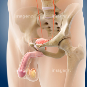
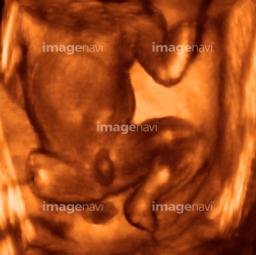
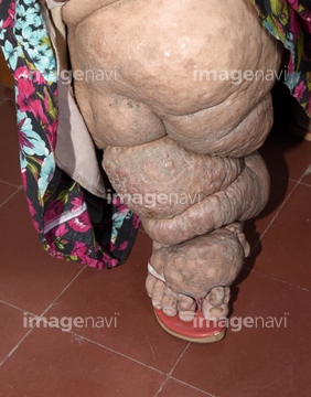
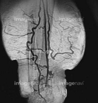
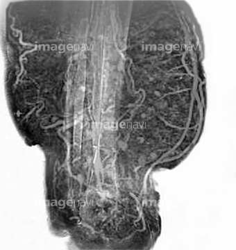
この検索結果には、Hydrocele、Foetal genitalia, 3-D ultrasound scan、Dwarfism, historical image、Swollen scrotum、Swollen testicles、Epididymo-orchitis from self catheterisationなどが含まれています。
64207190
64207191
64179887
64222716
64264687
64168089
64188193
64188391
64188392
64190960
64222705
64222706
64222707
64167749
64264805
64222360
64179884
64125400
64264623
64173392
64222361
64037924
64135634
64135635
64021719
64021720
64222359
64222708
64222710
64179881
64179890
64173394
64264688
64140719
64140720
64152321
64244403
64125421
64264761
64264765
64189204
64222709
64222711
64222712
64179880
64011971
64173395
64206147
64140721
64186259
64188276
64204477
64200829
64179882
64189201
64135638
64135645
64135654
64135659
64222779
64222780
64125422
64125423
64038905
64057701
64057702
64057703
64057704
64057705
64057706
64057707
64057708
64057709
64057710
64022005
64122845
64122846
64264693
64252686
64225827
64065583
64038757
64152598
64188411
64189202
64189203

64112463
64049854
64182480
64182494
64173494
64179885
64264644
20548478
20548493
20521289
64264642
64264643
64182459
64041678
64188255
64152551
64140722
64152302
64256447
64262998
64263045
64263250
64190948
64253262
64253263
64253264
64264770
64190940
64204337
64204338
64189208
64158564
64248414
64169570
64169571
64135636
64135637
64135639
64135640
64135642
64135644
64135655
64135658
64135660
64135661
64135663
64152279
64151275
64151699
64221714

64221716
64221718
64221734
64221739
64225826
64073957
20529186
17200053
17201294
64085238
64222788
64111602
64111625
64038950
64173983
64182590

64179883
64065614
64065775
64092844
64073958
20581977
20581982
20582070
20582073
64159466
53122348
64086307
20582397
| 次ページ |
