HOME > 写真 > 医療・福祉 > 医療 > 医療イメージ
10,000件の写真素材が検索されました。
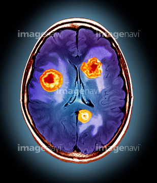
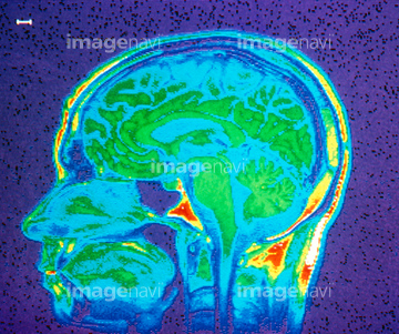
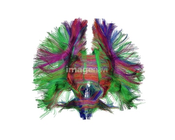
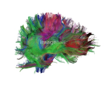
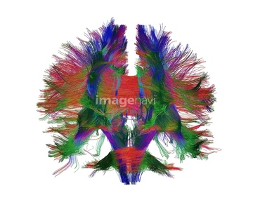
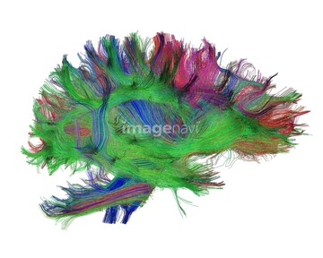
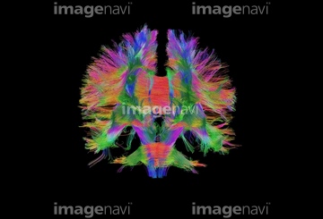
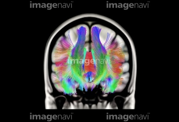
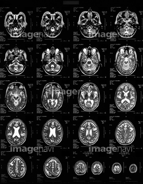
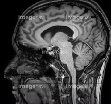
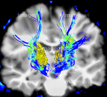
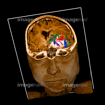
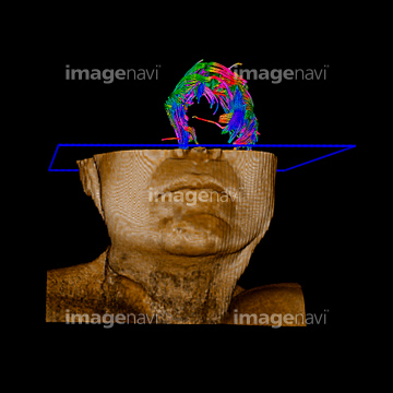
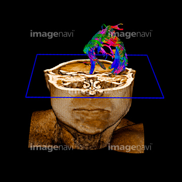
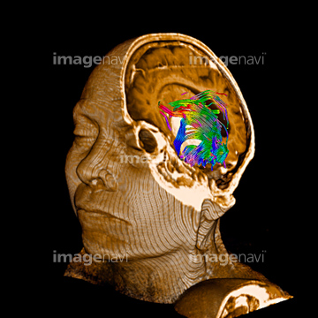
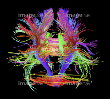
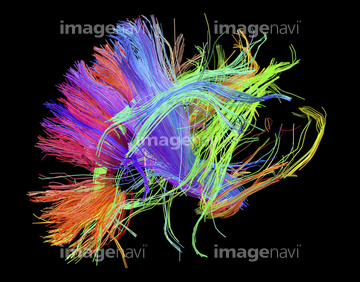
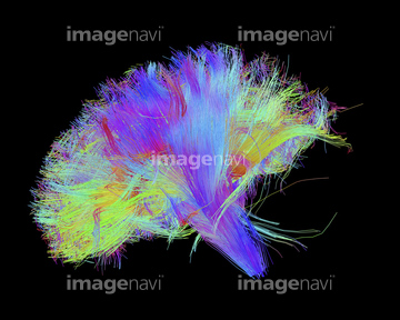
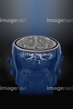
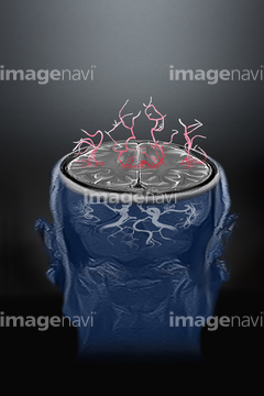
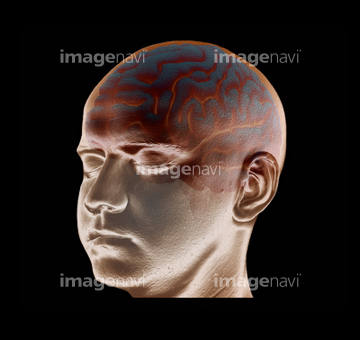
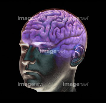
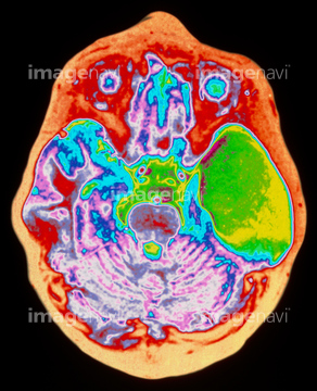
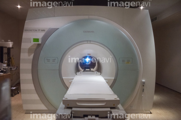
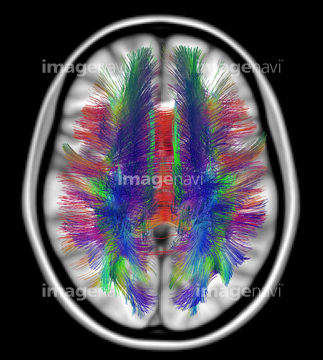
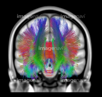
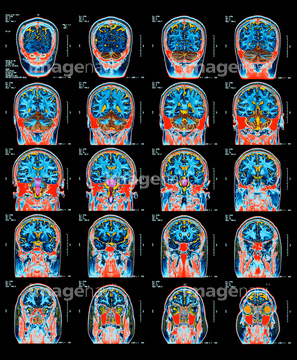
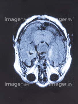
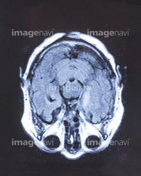
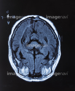
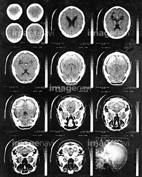
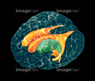
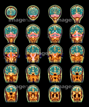
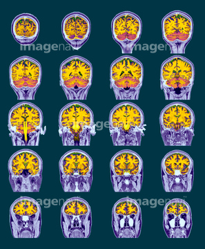
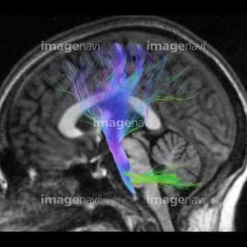
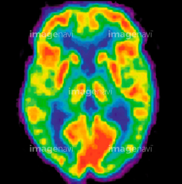
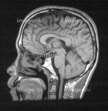
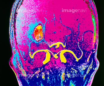
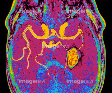
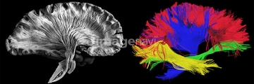
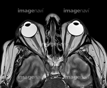
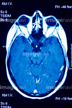
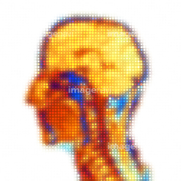
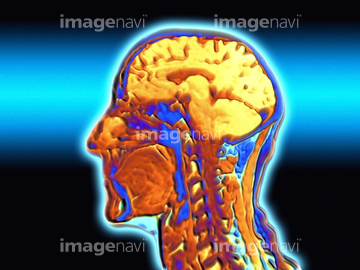
この検索結果には、Brain tumour, DTI and MRI scans、White matter fibres of the human brain、White matter fibres, brain mri scan、MRI scans、Brain MRI、MRI scan of man's head, side view (Digital Composi…などが含まれています。
64202762
64043131
20582138
20582139
20582140
20582141
20582142
20582143
20582144
20582145
20582146
20582147
20582148
30342237
64219589
64049886
17201401
30410250
64051975
64049872
64049873
64075285
64161171
64161172
64161174
64161176
64161177
64161178
64161182
64161187
64161200
64161201
64161204
64043125
64043128
64043129
64043130
64075025
64075026
64075027
64075028
64060094
64060095
10941464
30383186
30383185
64021473
64046179
64064693
30410251
30410253
64071706
64071707
64110133
64172494
64172495
64172496
64161164
64161165
21508369
64071942
64066826
64066827
64066833
64066834
64066835
64187413
64021451
64021452
64021453
64246618
17201404
17280779
17280780
17280781
17280783
17280784
17280785
30342233
30342234
64021454
64021468
64021469
64021470
64021471
64021472
64021478
64071943
64220127
64220128
64220129
20993281
20500112
64014403
64021456
64064694
64040391
64040392
64163476
64130747
17260856
16918314
16918315
64021447
64042984
64059701
64046178
64071925
64071927
64071447
64161179
64161181
64161183
64161194
64161202
64114171

64151747
64246712
64188464
30064857
64071453
17218208
64021475
64021477
64021479
17218188
17218191
| 次ページ |
