HOME > 写真 > 科学・テクノロジー > 科学 > DNA・細胞
10,000件の写真素材が検索されました。
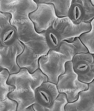
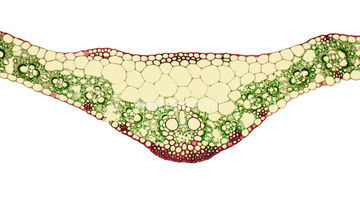
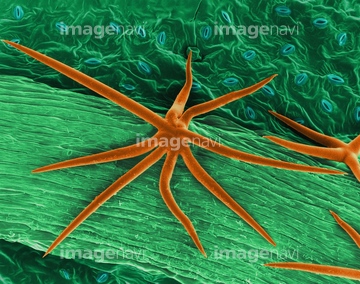
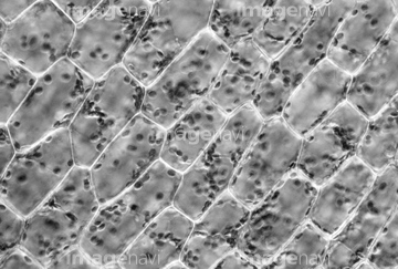
この検索結果には、Oak Leaf Stoma (Quercus robur)、Copper Beech Leaf Stoma (Fagus sylvatica)、Lavender leaf SEM、Pelargonium leaf, SEM、Open and closed stomates on leaf surface、Plant leaf trichome (Hibiscus sp.), SEMなどが含まれています。

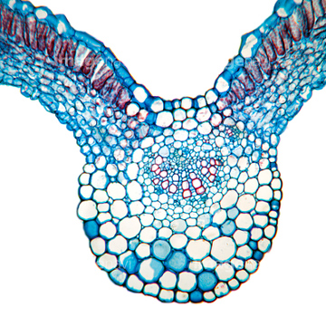
64210424

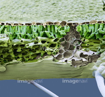
64005663
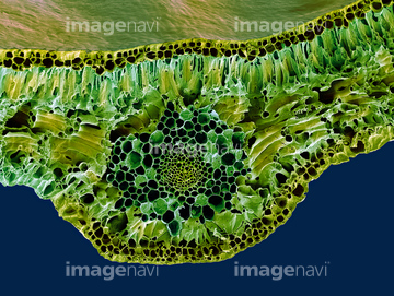
64005731
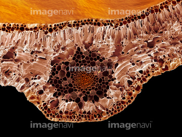
64005706
64005726
64005727
64083998
64083993
64083997
64012409
64005728
64005729
64005704
64005705
64005703
64005662
64075216
64083992
64083994
64083995
64083996
64012431
64012403
64004043
64005683
64005684
20542488
64005660
64005641

64087657
64087662
64005622
64005688
64259971

64064827
64005630
64005631
64005633
64005699
64151007
64005732
64097172
64005642
64005643
64005644
64005648
64005638
64005665
64005666
64005675
64005689
64005765
64005766
64005709
64087655
64076443

64004044
64005761
64005762
64005763
64250117
64137288
64137289
64137290
64137291
64087493
64137210
64150985

64114192
64124825
64124833
64005677
64114726
64005629
64005632
64005657
64005707
64098601
64260028
64260030
64098582
64098583
64098585
64208905
64208906
64208907
64144821
64048348
64250239
64250242
64250244
64250575
64250581
64250582
64157866
64056494
64005640
64089520
64089521
64107085
64107092
64005627
64005628
64005637
64005681
64005682
64005716
64005717
64005722
64148624
64005697
64126577
64002864
| 次ページ |
