HOME > 写真 > 医療・福祉 > 医療 > 手術
10,000件の写真素材が検索されました。
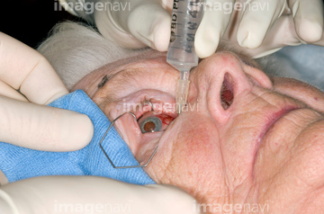
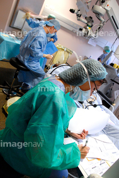
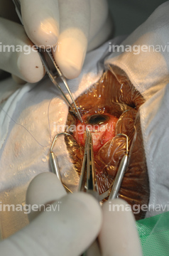
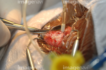
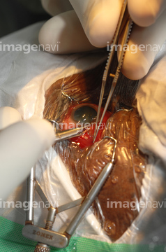
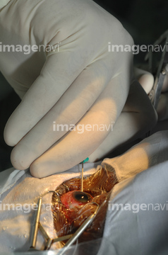
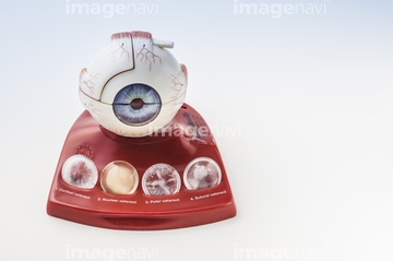
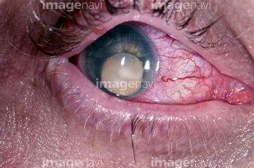
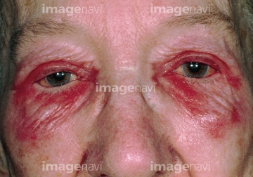
この検索結果には、Laser eye surgery、Lanosterol molecule、Optics of near-sightedness、Allergic conjunctivitis、Eye surgery、Spinal decompression surgeryなどが含まれています。
64222036
64222034
64222035
64012289
64012290
64222857
64012292
64012293
64019674
64012295
64012297
64173511
64222017
64222018
64243092
64012296
64012291
64010643
64010644
64010645
64012294
64243091
64243100
64243350
64243354
64224784
64102367
64102377
64019673
20503343
20503332
20503333
20503334
20503339

20503340
64008559
64237457
64237458
64188994
64243391
64168147
64168148
64008562
17256381
64107769
64264814
20536861
20536862
20536863
20536864
20536865
20536866

20536867
20536868
64214325
64222718

17200431
64168146
64190956
64214319

20503321
20503322
20503323

20503324
20560380
64011580
20564713
20564731
64222945
64222946
64040291
64222661
64222662
64255242
64182588
20503330
20503331
20503335
20503337
20503338
20503326
64242742
64264701
64219641
64222245
64222246
64222247

20549421

20549422
64247753
64247754
64222021
64222022
64008485
64037783
20531001
20531002
20531003
20531004
20531005
20531006
64151407
64107580

20562951
64188311
64188350
64188351
64188353
64165710
64165711
64165712
64047479
64047480
64217943
64222215
64222216
64222217
64222218
64222219
64060379
64060381
64214331
31783503
20582372
20582373
20582374

64244333
64223413
64056992
64056995
64056996
64056999
64157285
64122891
64014084
64242418
64158721
64158722
64115855
64019672
20503341

20503342
64213683
64264819
64173516
64173517
64114190
64102363
64102381
64151405
64153741
64153742
64201377
64201378
64201380
64109011
64011573
64011574


64217941
64217942
64172411
20560452
64222644
64222647
64222648
64222649
| 次ページ |
