HOME > 写真 > 医療・福祉 > 医療 > 手術
10,000件の写真素材が検索されました。
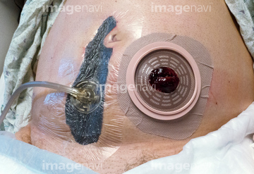
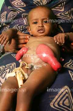
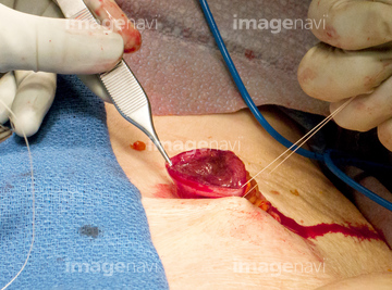
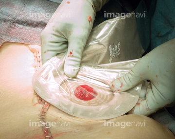
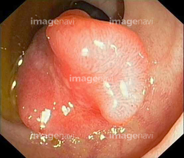
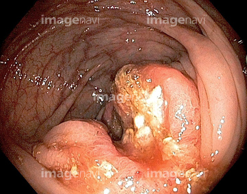
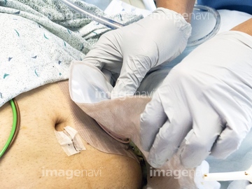
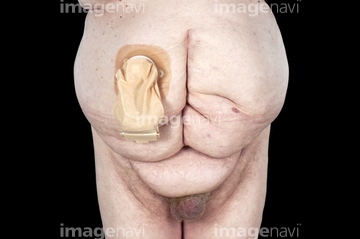
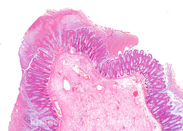
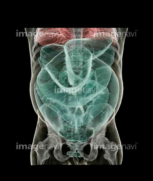
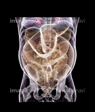
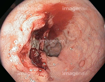
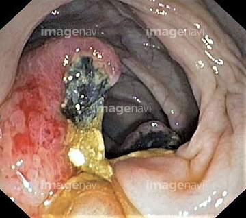
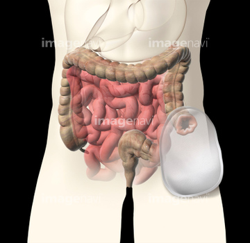
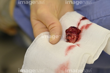
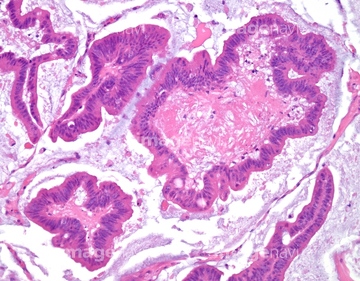
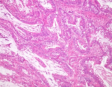
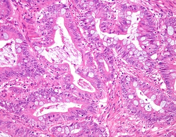
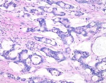
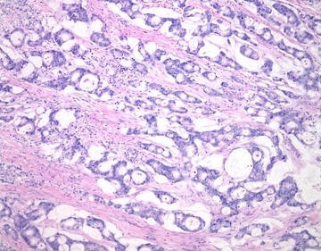
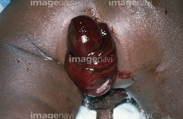
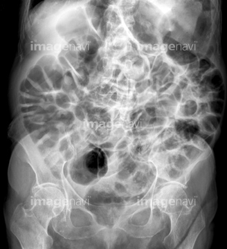
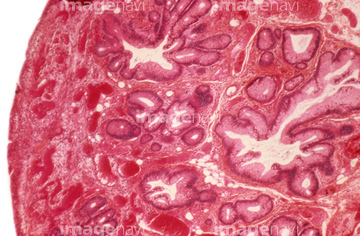
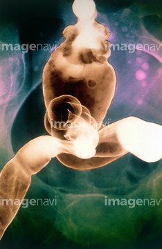
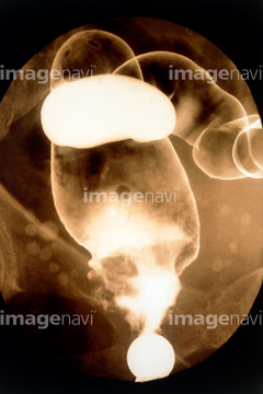
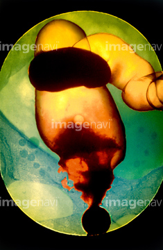
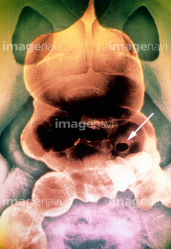
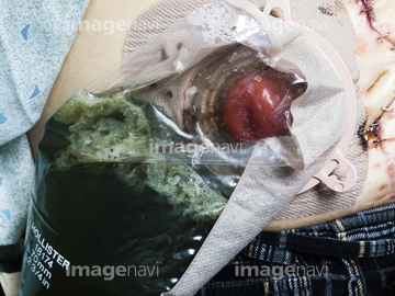
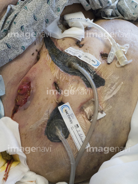
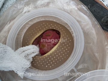
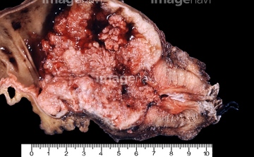
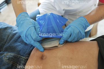
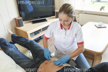
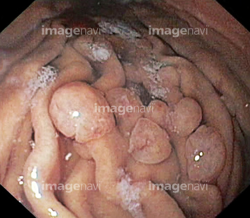
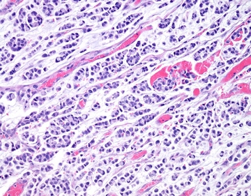
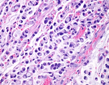
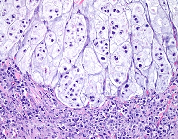
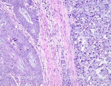
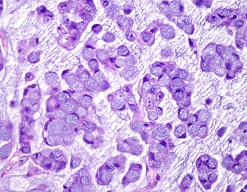
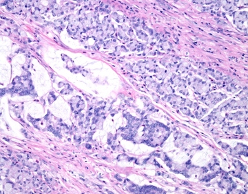
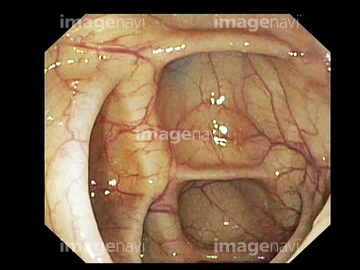
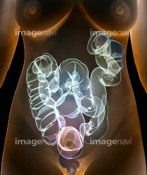
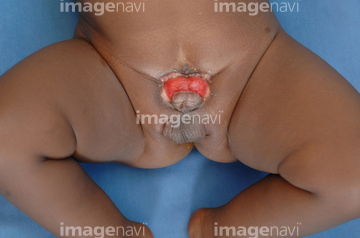
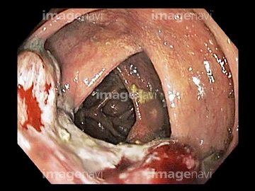
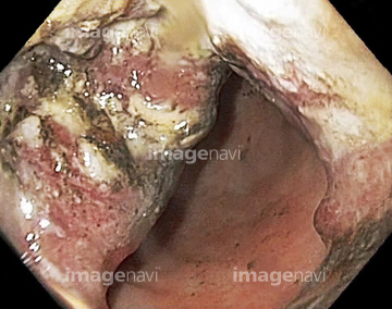
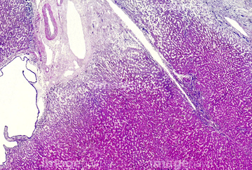
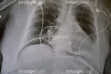
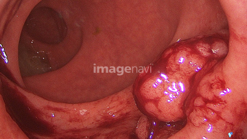
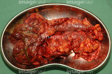
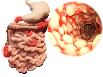
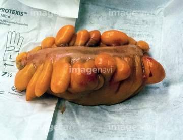
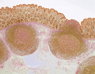
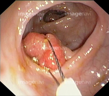
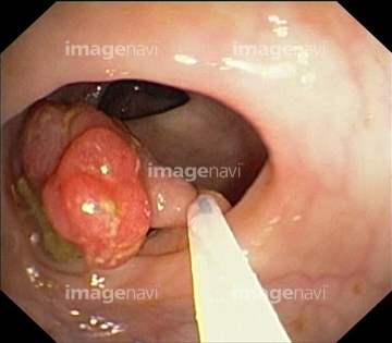
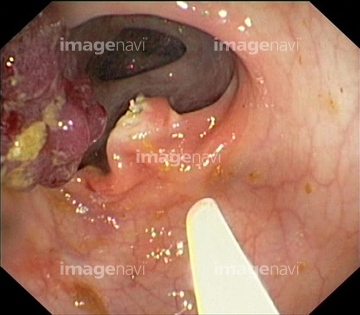
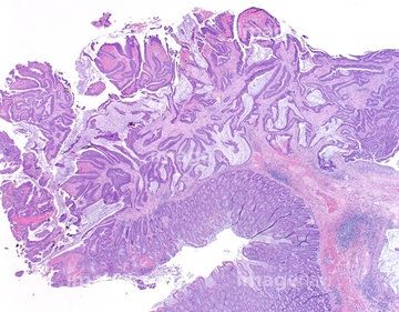
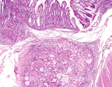
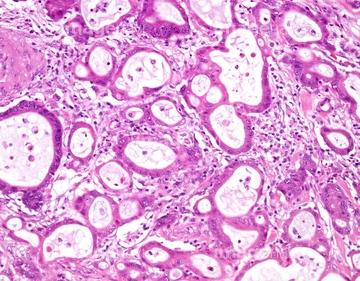
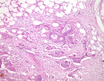
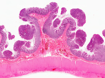
この検索結果には、Colostomy bag and intestines, artwork、Excised polyp、Ostomy bag, clip and adhesive wafer、Mucinous carcinoma of the colon, light micrograph、Intussusception of the colon、Abdominal blockage, X-rayなどが含まれています。
64222811
64123601
64135615
64135666
64131737
64123606
64126635
64135633
64222774
64123624
64207283
64125442
64125443
64125571
64135616
64170398
64170400
64170401
64170403
64170404
64170405
64170406
64170407
64170408
64170409
64170410
64170411
64170412
64125444
64130017
64130018
64125570
64123603
64170430
64126650
64090184
64170399
64170402
64174395
64188419
64157255
64157256
64157257
64259895
20559007
64264733
64099577
64222805
64123592
64205896
64205907
64088333
64088334
64088335
64088337
64128087
64135608
64135626
64135629
64219686
64219687
64219688
64219689
64157259
64157260
64158641
64125823
64125574
64071846
64071847
64077208
64222812
64176633
64176636
64176639
64088768
64088770
64089598
64088310
64122849
64122864
64173555
64213803
64128103
64259839
64222220
17235927
64129306
64207220
64207279
64135617
64135618
64088340
64139024
64184356
64184357
64157258
64165820
64165821
64196498
64208387
64222673
64222674
64055695
64225318
64222775
64155657
64155701
64184343
64088332
64088338
64088341

20559280
64125573
20531320
20531321
20531322
64123691
64123692
64184354
64184355
64259876
64263851
17234346
17256490
17256491
17256492
17256493
64063349
64075507
64264698
64264823
20531370
64168095
64253238
64078994
64250036
64222341
| 次ページ |
