HOME > 写真 > 医療・福祉 > 医療 > 医療器具
10,000件の写真素材が検索されました。
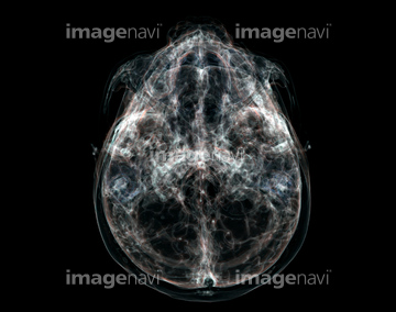
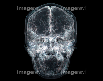
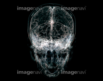
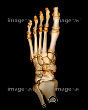
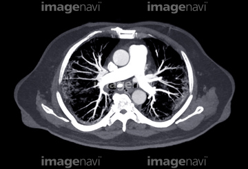
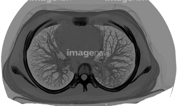
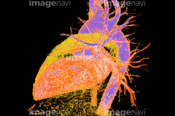
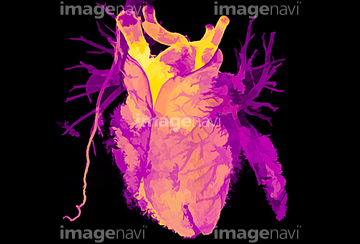
この検索結果には、Bronchiectasis, 3D CT scan、Kommerell diverticulum, 3D CT scan、CT scanning、Thorax, CT scans、Urography、Human skull, CT scanなどが含まれています。
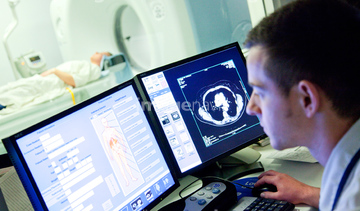
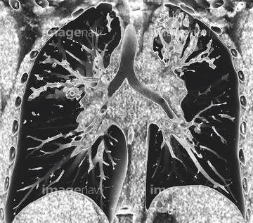
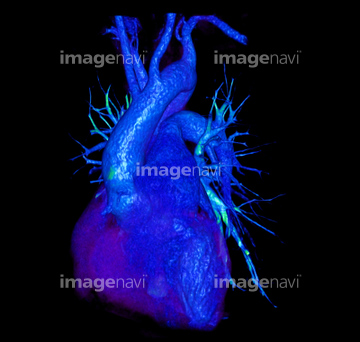
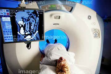
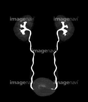
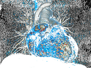
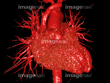
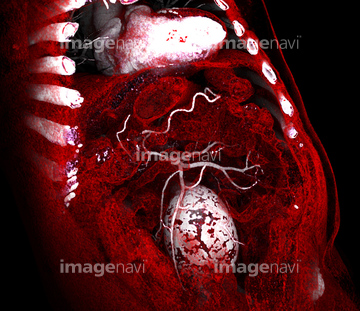
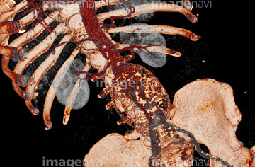
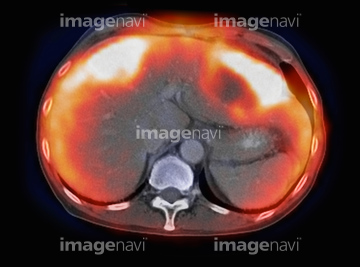
64223185
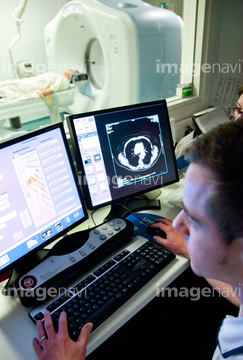
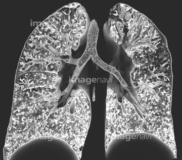
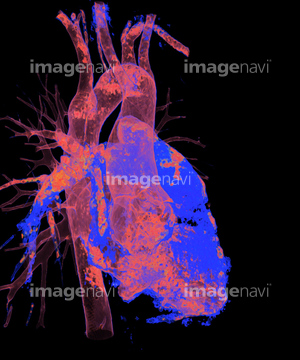
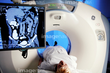
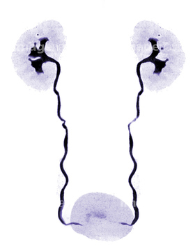
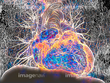
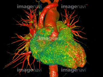
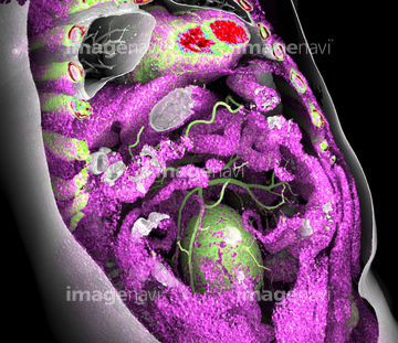
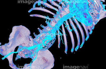
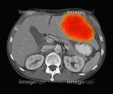
64223186
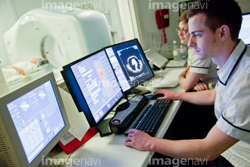
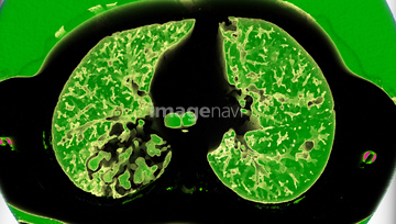
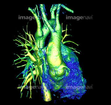
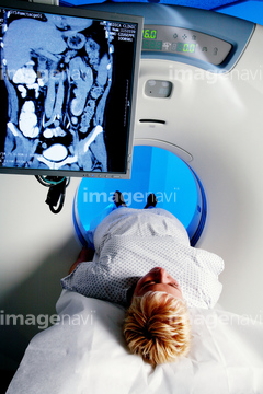
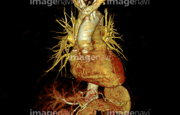
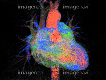
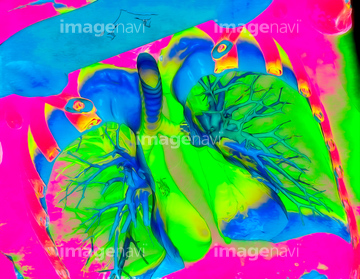
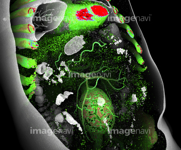
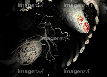
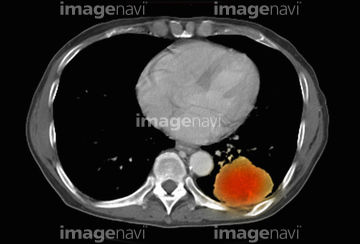
64222838
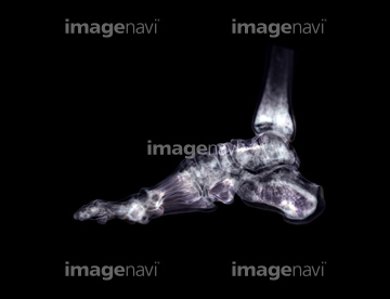
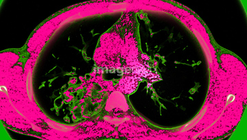
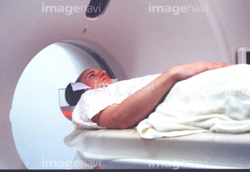
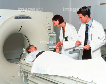
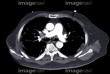
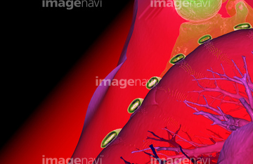
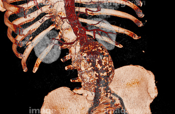
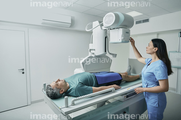
20569532
20569533
20569534
20569535
20569536
20569537
20569538
64223194
64242452
64057026
64057027
64057028
20569549
20566904
20569523
64037563
64019120
20555751
20555785
64209042
20569526
20569550
20569551
20569513
20569517
20569515
20569516
20569518
20569519
20569521
20569522
20569544
20569545
20569547
20555796
64248809
64248810
64248811
64248812
64248813
64248814
64248815
64248816
64248817
64248818
64248819
64248820
64248894
64248901
64248902
64248903
64019118
64019119
20562185
64019145
20569561
64130468
20569520
20569546
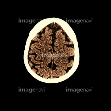
64222731
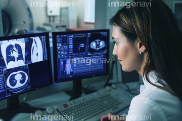
64052088
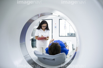
64087003
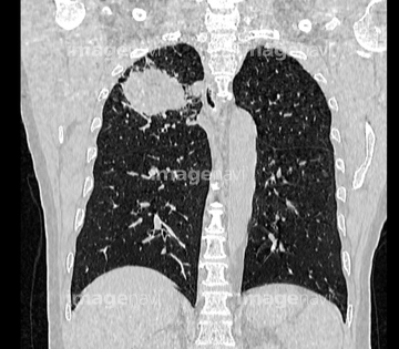
64021288
64021289
64250825
20567594
20573161
20564379

17246303
20573133
20573134

20573135
20573136
20573137
20573138
20573139
20573140
20573141
20555739
64243526
64019133
64019134
64019135
64019116
64021322
64021323
64019127
64019128
20569543
20569555
20569557
20569559
64019137
64220195
20569525
20567664
64087911
64226421
17238388
20573151
20573152
20573153
20573154
20573155
20573156

20573157
20573627
20573629
20573632
20573633
20573663
20573690
20573691
20562152

64257681
64257682
64257683
64257684
64257685
64257686
64257687
64257688
64257689
64257690
64257691
64257692
64257693
64257694
64257698
64257699
64257734
64257735
64257736
64257741
64257744
64257745
64257746
64257747
64257748
64257768
64257784
64157634
20562680
17271015
20555798
20543695
64203174
64203175
64203176
64203177
64203178
64203179
64203180
64203181
64221278
64223091
64248830
64248831
64248832
64248833
64199310
64138928
64138929
20555795
64248802
64248803
64248804
64248805
64248806
64248807
64248808
20567528
10315262
64110144
17200589
64245663
64245665
64189038
64243361
| 次ページ |
