HOME > 写真 > 医療・福祉 > 医療 > 病院・クリニック
10,000件の写真素材が検索されました。
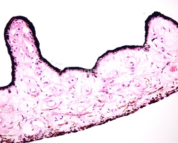
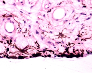
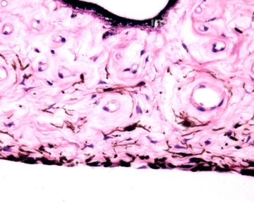
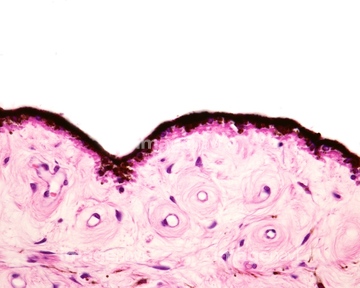
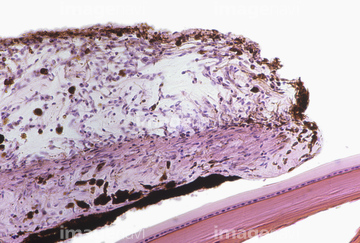
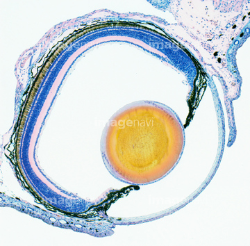
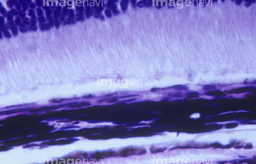
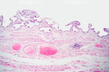
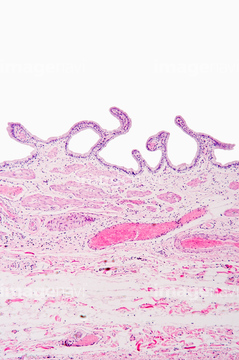
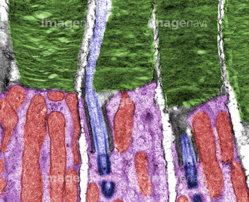
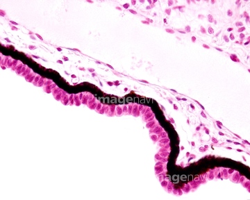
この検索結果には、Blood vessels in iris stroma, light micrograph、Iris pigment cells, light micrograph、Iris layers, light micrograph、Anterior surface of iris, light micrograph、Iris, light micrograph、Cornea, light micrographなどが含まれています。
64224839
64190721
64190723
64190724
64190726
64225221
64260274
64216438
64216441
64216442
64056270
64198977
64198978
64114522
64114523
64114524
64260017
64198974
64216424
64260021
64090168
64090170

64260018
64190713
64194306
64216423
64216439
64216440
64224841
17235966
64145910
64093922
64198945
64115571
64190681
64190682
64190689
64190690
64190691
64190692
64190693
64190696
64190709
64190710
64190712
64198955
64198956
64198958
64198959
64198963
64198964
64198966
64198967
64198968
64198969
64198973
64216434
64216435
64213935
64225213
64225212
64225132
64096078
64114570
64114571
64260020
20532901
64148801
64148804
64225224
64114521
64198948
64198960
64198961
64198962
64088778
64115581
64114540
64224844

64114549

64224928
64225231
64263134
64190680
64194261
64194262
64194263
64194264
64194271
64194272
64194273
64194295
64194296
64194297
64194299
64194300
64194301
64194303
64194338
64194340
64194341
64194352
64194353
64197461
64205315
64205318
64205319
64207667
64213894
64213930
64114577
64114561
64235075
64235076
| 次ページ |
