HOME > 写真 > 人物 > 構図 > 色バック
10,000件の写真素材が検索されました。
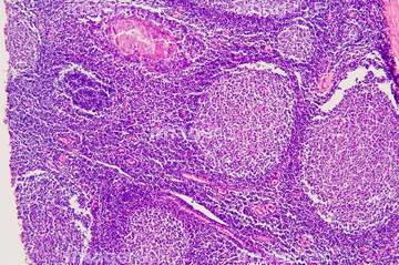
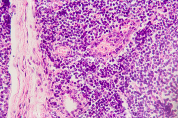
この検索結果には、Sarcoma, TEM、Heart valve and strings, SEM、Contracted heart muscle fibre and capillary, SEM、Hodgkin's lymphoma, light micrograph、Vein cross-section. LM、Sarcoma cancer cell culture, SEMなどが含まれています。
64225245
64224908
64166185
64116650
64116651
64247940
64123989
64205895
64073994
64073995
64166157
64166160
64224909
17200418
64244160
64244206
64155658
64161283
64161297
64161298
64247921
64247922
64247923
64247947
64247948
64178675
64178680
64073996
17212300
64115571
64008145
64008147
64224951
64224954
64150221
64132349
64132350
64039248
64190702
64190703
64190704
64247882
64247917
64247924
64247925
64247926
64247927
64225236
64192928
64073997
64257220
64257221
64257222

64224953
64225232
64225233
64225293
17212277
17212278
17212293
17212294
17212301
17212302
17212310
17212311
64096074
64096084
64150358
64133042
64133043
64224929
64256046
64256047
64256048
64256049
64256050
64256051
64256052
64256053
64224932

64224937
64224938
64224950
64224904
64224907
64224911
64225231
20530811
20530977
20531037
64224927
64225204
64197474
64245184
64245187
| 次ページ |
