HOME > 写真 > 自然・風景 > 大地 > 台地
10,000件の写真素材が検索されました。
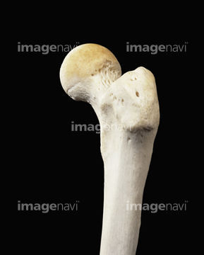
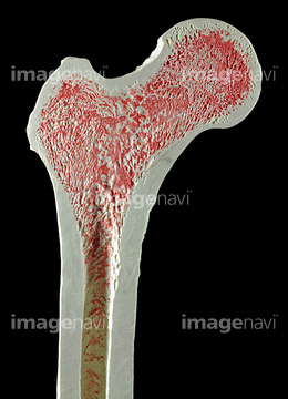
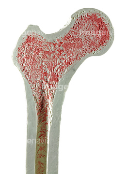
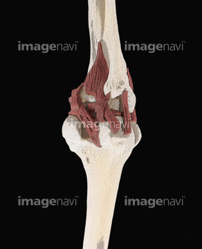
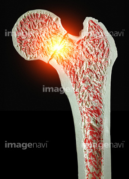
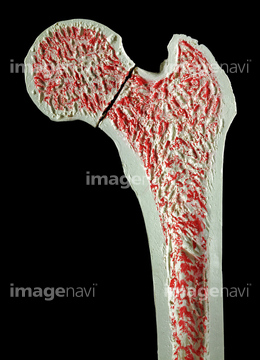
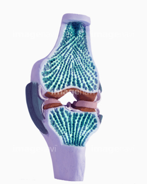
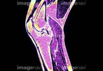
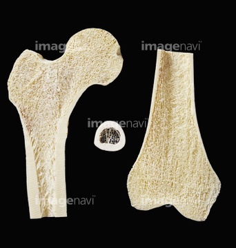
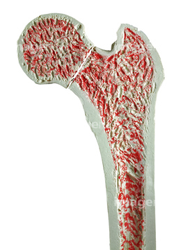
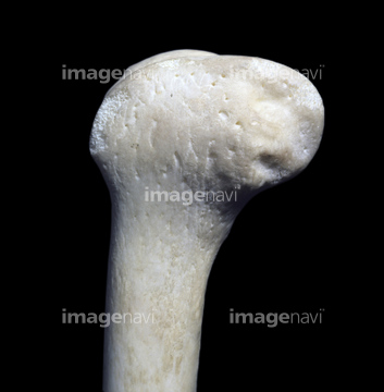
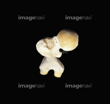
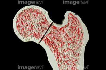
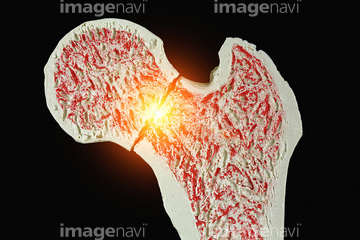
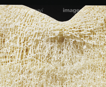
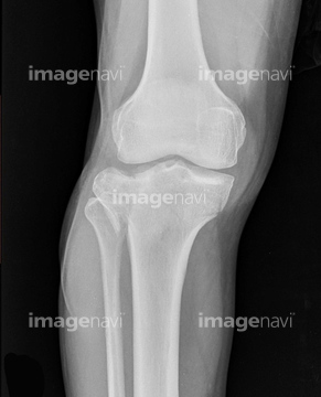
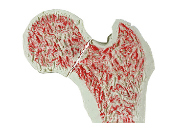
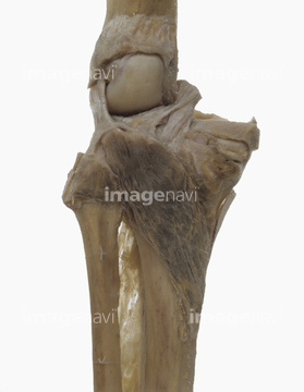
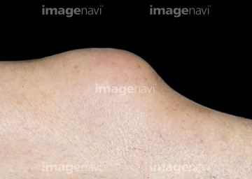
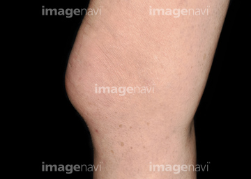
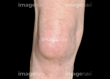
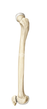
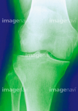
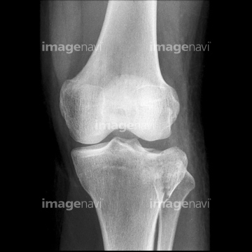
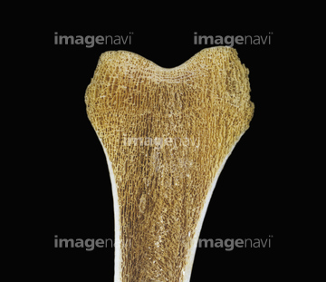
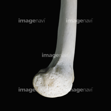
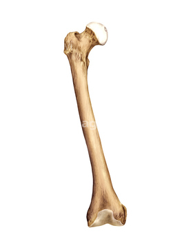
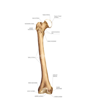
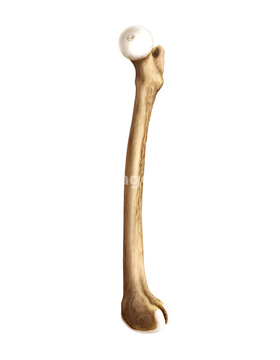
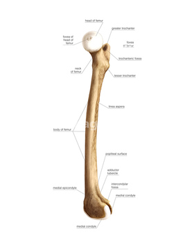
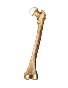
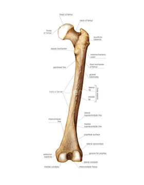
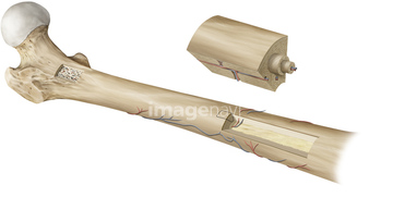
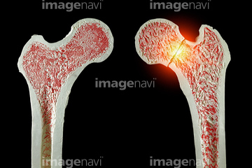
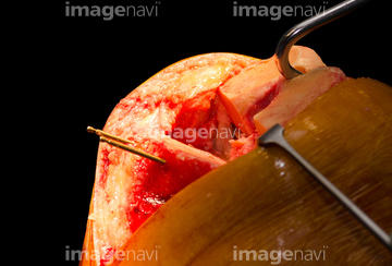
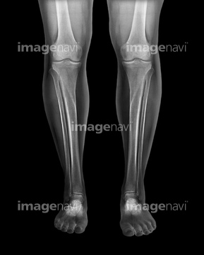
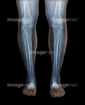
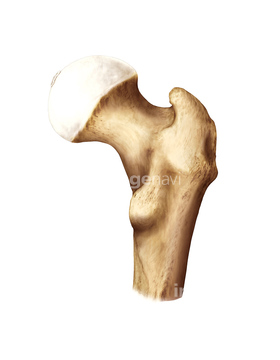
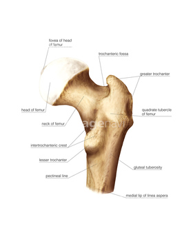
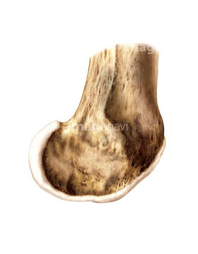
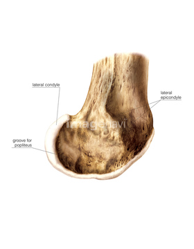
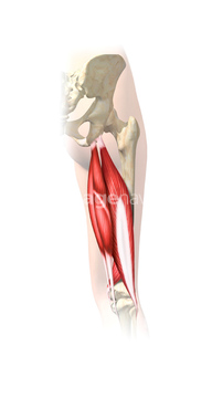
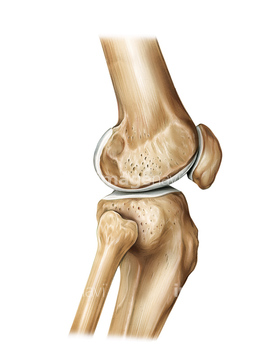
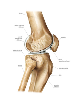
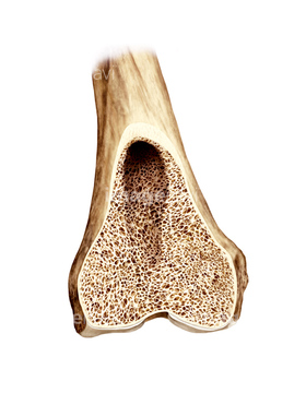
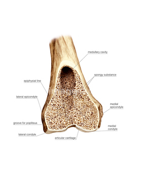
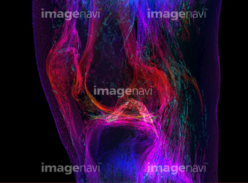
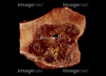
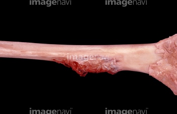
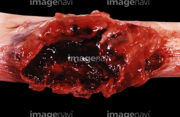
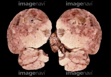
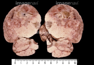
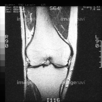
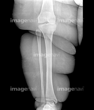
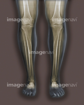
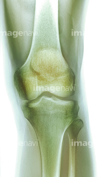
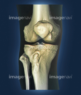
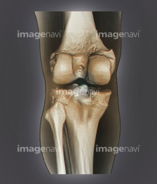
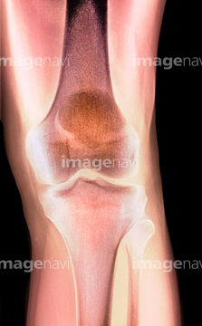
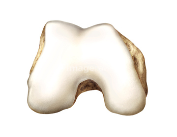
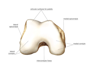
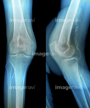
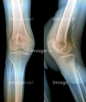
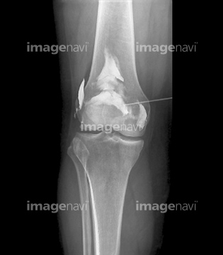
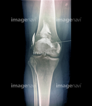
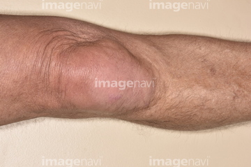
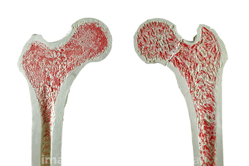
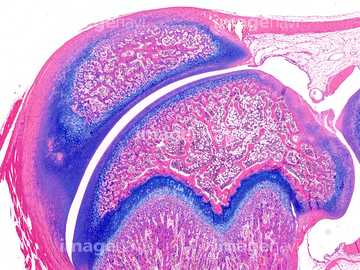
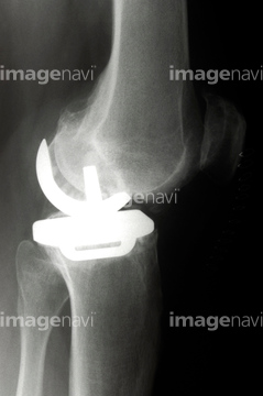
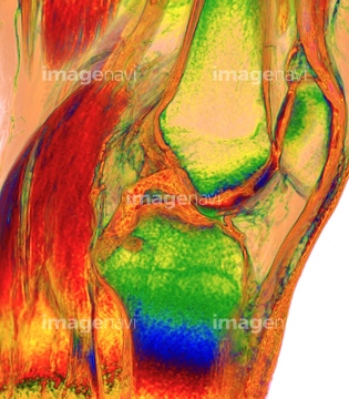
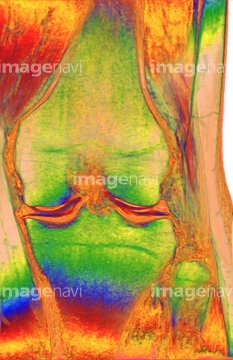
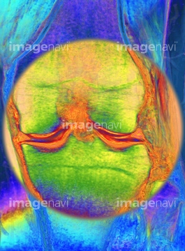
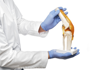
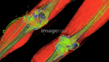
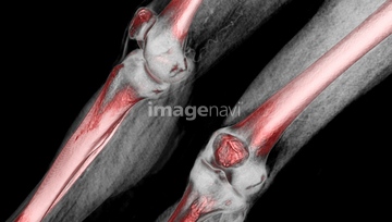
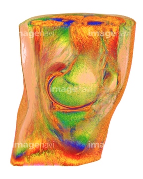
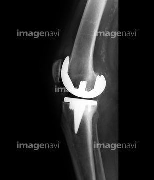
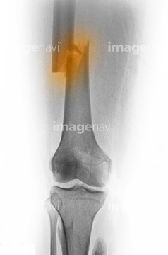
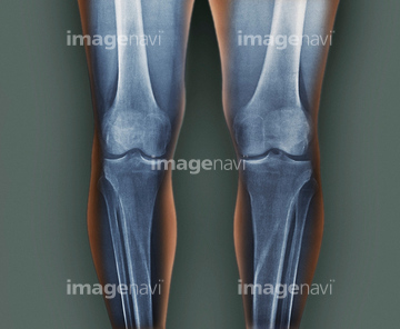
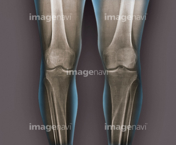
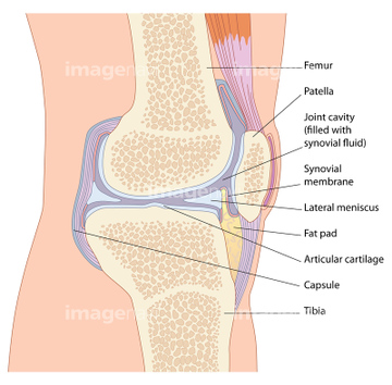
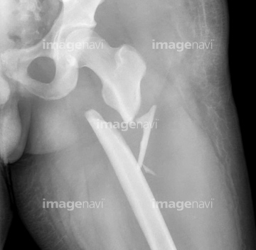
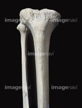
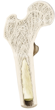
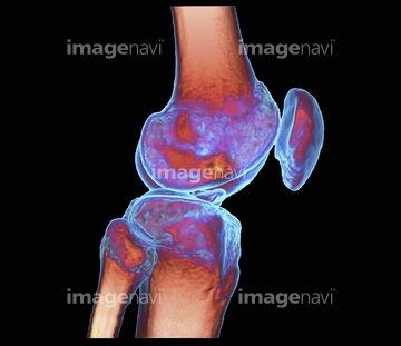
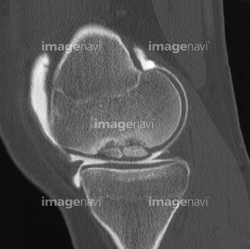
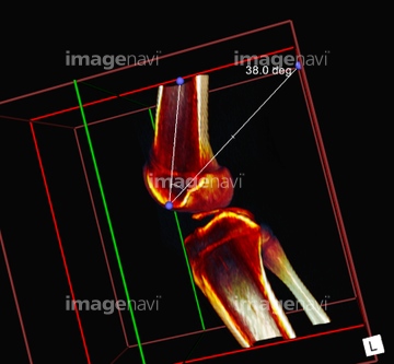
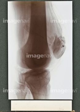
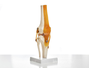
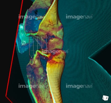
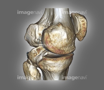
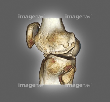
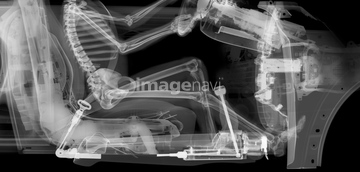
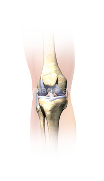
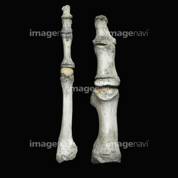
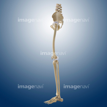
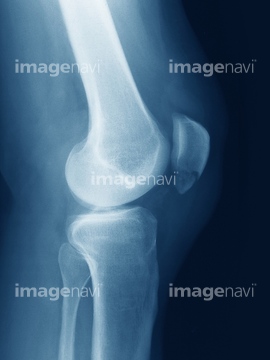
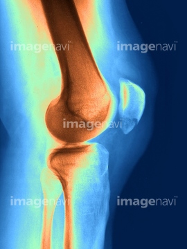
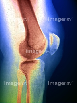
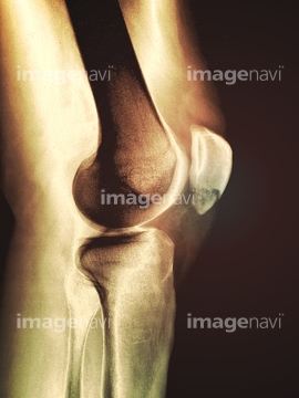
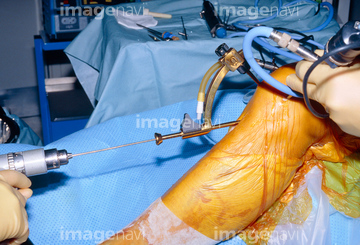
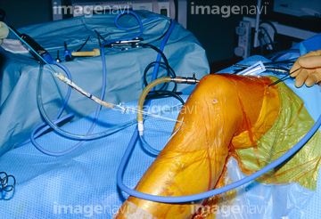
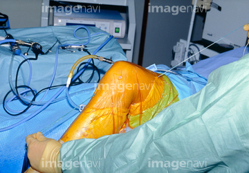
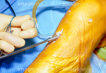
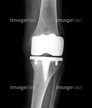
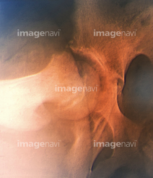
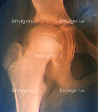
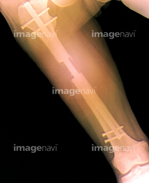
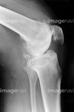
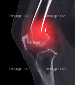
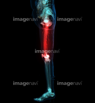
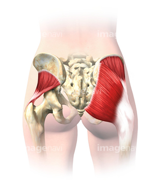
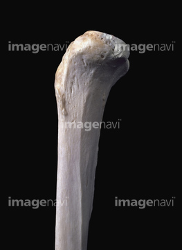
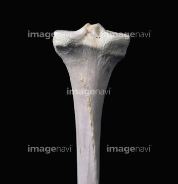
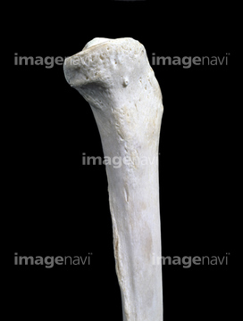
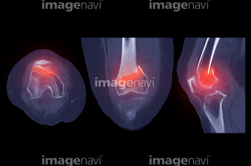
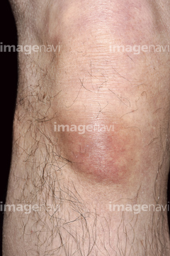
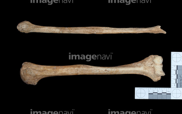
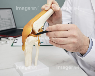
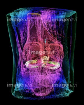
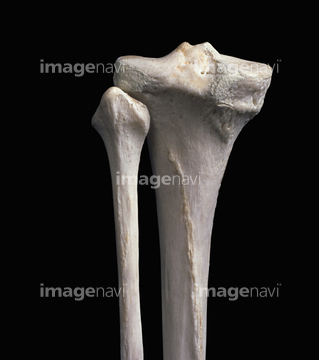
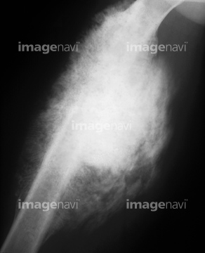
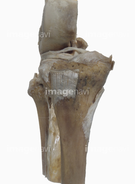
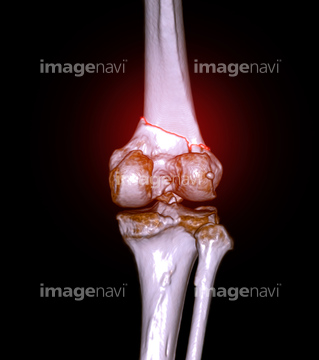
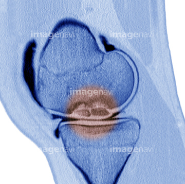
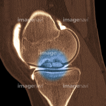
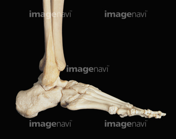
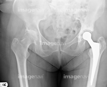
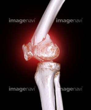
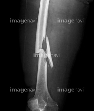
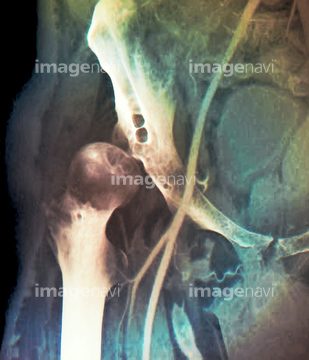
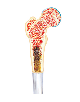
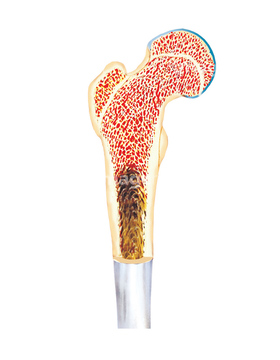
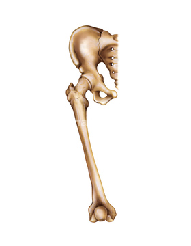
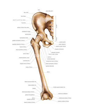
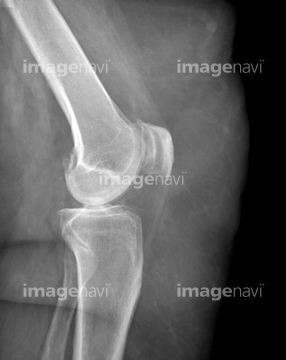
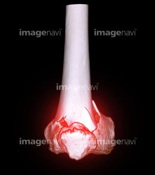
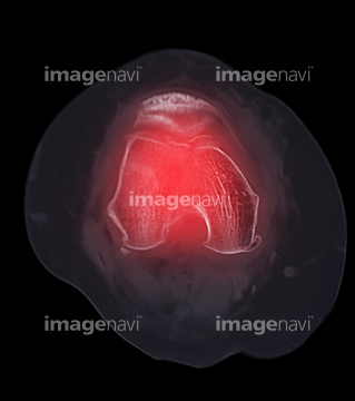
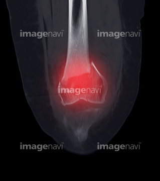
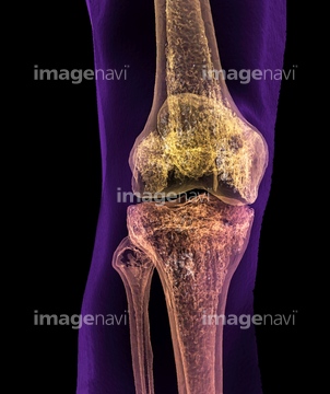
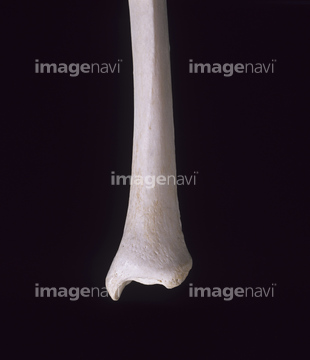
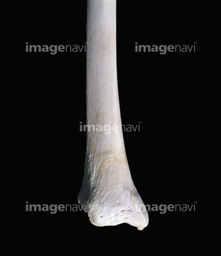
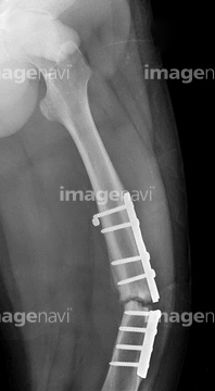
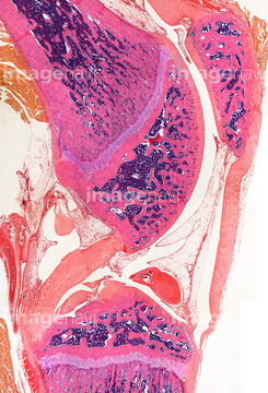
この検索結果には、Normal and fractured hip、Knee replacement surgery、Normal legs, X-rays、Proximal extremity of femur, artwork、Distal extremity of femur, artwork、Hamstrings, artworkなどが含まれています。
64225007
64225455
64224980
64225006
64225382
17237153
17237147
64225117
64225369

64225468
64225391
64225397
17237144
17237158
64225118
17237156
64225469
17237145
17237155
64225003
20559314
20559315
20559316
20559317
20559318
20559319
20559320
20559321
20559322
17237149
64225370
64174369
64174370
64174371
53122342
17201772
20559313
64224999
64225298
64070807
64070808
64070809
64070810
64070811
64070812
53122649
17237154
64059505
64064639
64064640
64070813
64070814
64070815
64070816
64071951
64070821
64070822
64187411
17200869
64044869
64044870
64219700
64219701
64219702
64219703
64219704
64219705
64219706
64219707
64219708
64010066
64064641
64040050
64071469
64071470
64074821
64070817
64070818
64012607
64012608
64012614
64012615
64188280
17237157
64250019
64012194
64149490
64149491
64149492
64149493
64149494
64149496
64071954
64208800
64208803
64149495
17200868
64064637
64064638
64071693
64071695
64071696
64049300
64049302
20555788
53122377
17200867
64050519
64198085
64198087
64198088
64262496
64109321
64126201
20548930
64109322
64198083
64198084
20527600
64071956
64056862
64221971
64221972
64221973
64221974
64245408
64245409
64245410
64245411
64013810
64013813
64013814
64013815
17200865

17200866
64049831
64049847
64049874
64009848
20573699
20569560
64071950
64225355
64225419
64225465
20573700
64168172
64100527
20549492
64262494
64014105
64048561
64224992
64225438
64044871
64225371
20573694
64262498
64262499
64225004
64012992
64049832
20569554
20569555
20569559
20573695
20558093
64012562
64070560
64070561
64070801
64070802
64159940
64148942
20573692
20573697
20573698
64101867

64225297
64225392
64093968
64093987
| 次ページ |
