HOME > 写真 > 人物 > 体のパーツ
10,000件の写真素材が検索されました。
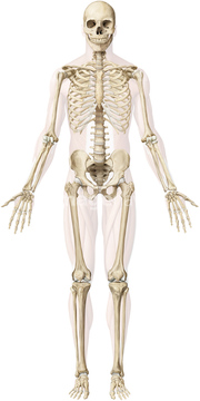
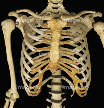
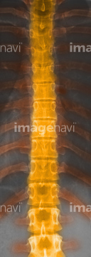
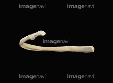
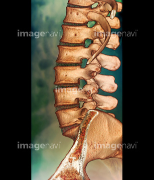
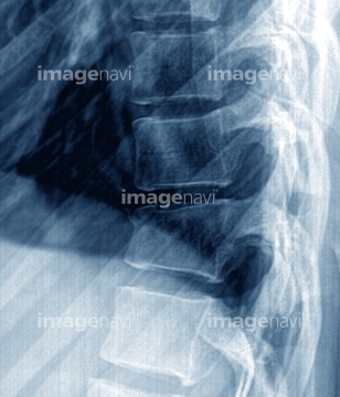
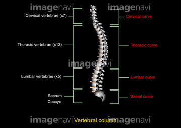
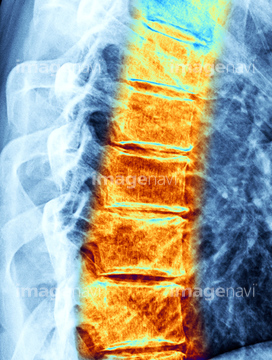
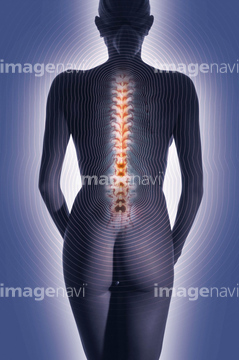
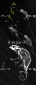
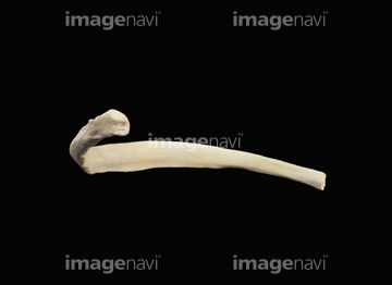
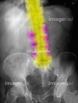
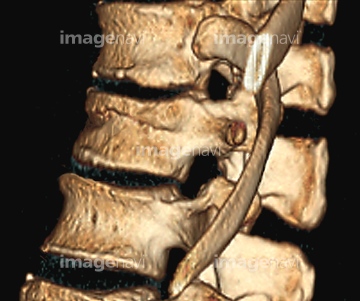
この検索結果には、Human spine, artwork、Upper limb bone、Bones of the sternum、spine x-ray、Right femur proximal end from behind、Healthy lower spine, X-rayなどが含まれています。
64225029
64225163
53122256
53122281

53122623
53122681
53122798
64225291
64225253
64225254
64225353
64225367
64225463
64225365
64224984
20572336
64225376
64225255
64225352
64225329
64225330
64225331
64225332
64225354
64225290
53122372
53122453
64220889
64225465
64225442
64225423
64021294
64225421
64225289
64225358
64225400
64225437
64225413
53122341
64225067
64239077
64225403
17212194
64225301
64225357
30328491
64225468

64021287
64225419
30094706
64225030
64225031
64225214
64225306
64225386
64225281
64220883
53122297
53122337
53122352
53122356
64225377
64225383
64225333
64225252
53122282
53122313
53122349
53122353
53122368
53122369
53122503
53122541
53122542
53122709
53122713
53122714
53122715
53122737
53122738
53122795
64225406
64225441
64225459
64225016
64225062
16934066
64225466
64225382
64225395
64225438
17237153
64225464
53122301
64225350
64225415
64225433
64225299
64225355
64225027
64227098
64225356
64225387
64225456
64225462
64119246
20530971
64225305
16934060
64225007
64225418
64225020
64263757
64224987
64225271
64225061
64225117
64225004
64225467
64224991
64225390
64224986
64224992
64225051
64225469
10995818
64225295
64225296
64225369
64225402
64225378
16934065
53122342
53122373
53122376
53122377

53122394
53122666
53122675
17205373
17205401
64225440
64225167
64220877
20571948
64225008
64225009
64225300
64048259
64077581
64013375
20571955

20571957
64221654
64221655
64224980
64040060
64225408
64225435

20571961
64012963
64040149
64040150
64225287
16934079
64225006
10919361
64225425
64260184
64225407
64220139
64225424
64240164
64224985
64225420
| 次ページ |
