HOME > 写真 > 乗り物・交通 > 海路・水路 > 運河
10,000件の写真素材が検索されました。
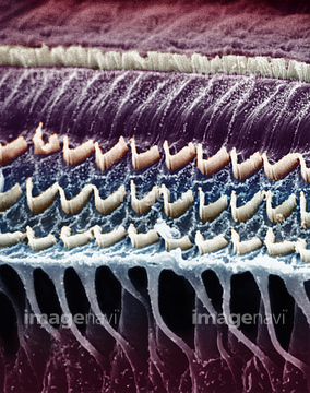
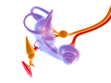
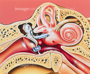
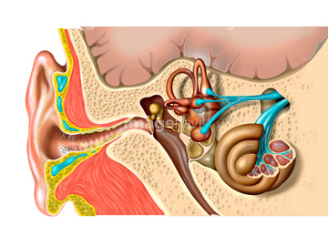
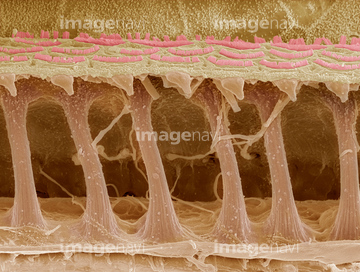
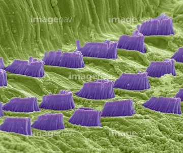
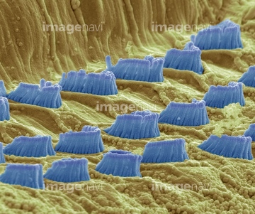
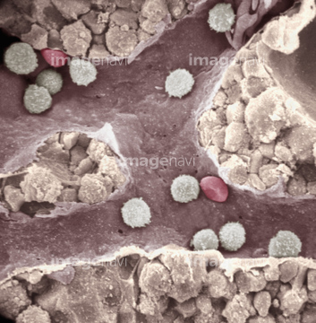
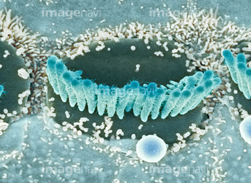
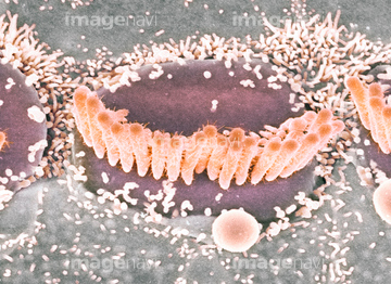
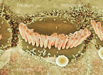
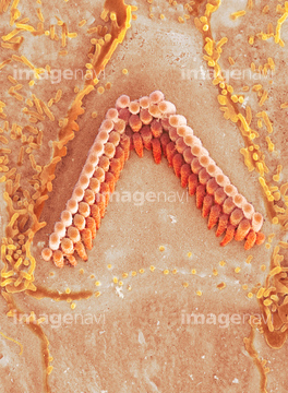
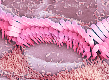
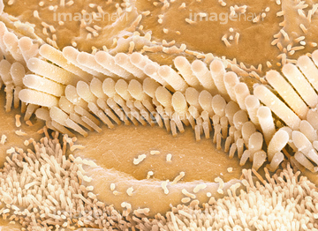
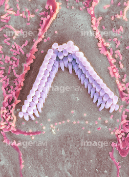
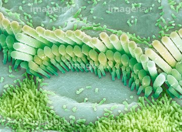
この検索結果には、Inner ear hair cells, SEM、Cochlea, light micrograph、Eye anatomy、Cryofracture preparation of sciatic nerve、Skin cross-section, SEM、Sea cucumber ossicles, SEMなどが含まれています。
64225197
64225195
64225069
64225070
64080566
64052109
64088778
64244789
64244828
64225063
64043308
64043311
64043314
64043315
64035023
64075654
64224897
64265183
64265184
64152604
64103881
64103906
64202541
64202542
64220308
64265185
64265186
64265187
64265188
64265189
64265190
64043309
64043312
64043313
64205071
64205072
64205073
64205074
64205075
64205076
64205077
64205078
64205079
64205080
64205081
64205082
64205083
64205084
64205085
64205086
64205087
64205088
64205089
64205090
64205091
64194305
64194351
17208462
64225132
64225159
64150935
64192851
64192881
64009067
64009071
64009072
64009074
64009076
64205141
64008675
64008677
64009051
64009056
64009063
64009064
64009065
64039926
64039927
64039928
64074757
64074758
64082470
64082471
64009170
64009171
64205059
64205060
64205061
64205062
64205063
64205064
64205065
64205066
64205067
64205068
64205147
64205148
64205149
64132047
64132048
64132058
64132065
64132080
64132088
64132091
64132093
64132100
64132101
64205092
64205093
64205094
64205095
64205096
64205097
64205098
64205099
64205100
64205101
64224892
17201417
64250856
64250857
64130489
64225176
64190166
64114169
64115471
64245072

64225083
64225088
64225129
64225130
64225131

64225133
64225151
64225152
64225172
64225190
64225192
64225198
64225201
64225202
64225261
64225361
64029976
64136970

64136971
64136972
64136973

64050571
64206768
64206769
64206770
64205315
64205316
64205317
64205318
64205319
64245206
64245207
64245208
64245221
64205069
64205070
64205133
64205134
64205135
64205136
64205137
| 次ページ |
