HOME > 写真 > 科学・テクノロジー > 科学 > DNA・細胞
10,000件の写真素材が検索されました。
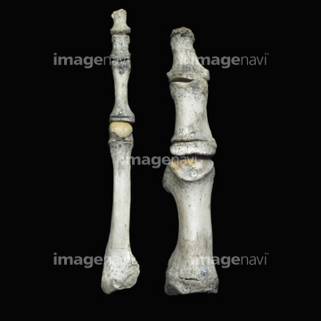
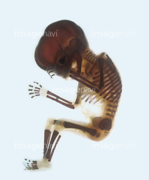
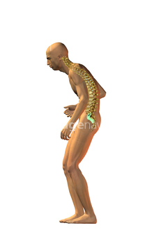
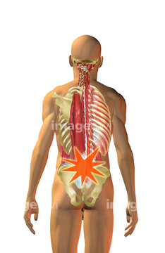
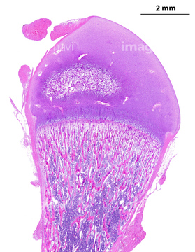
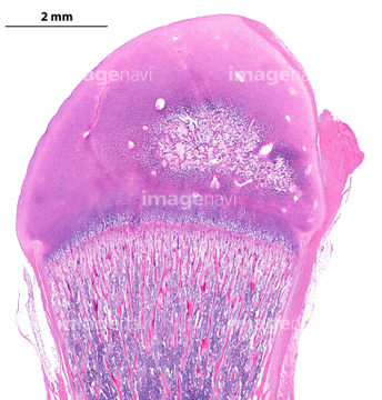
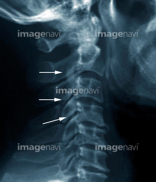
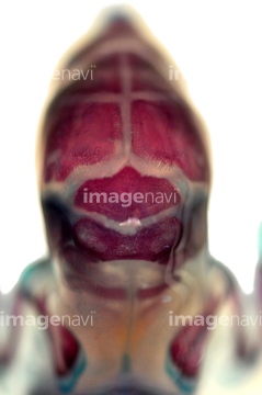
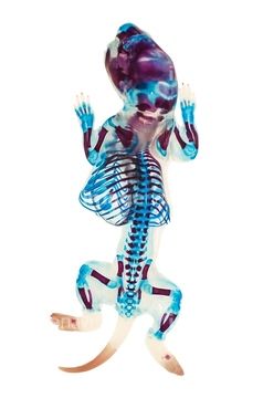
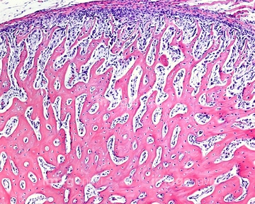
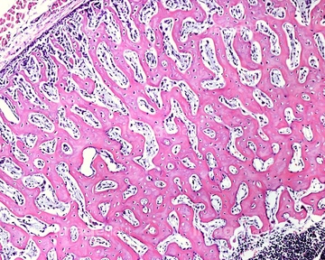
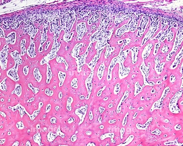
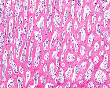
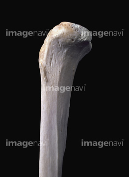
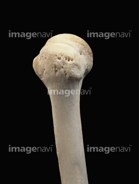
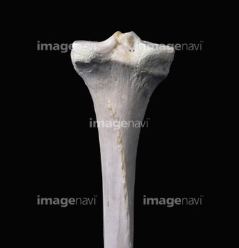
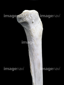
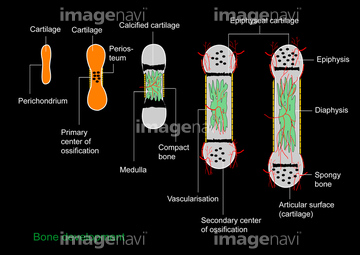
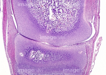
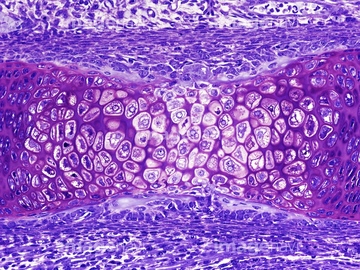
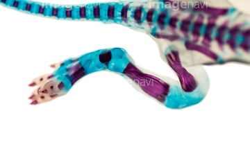
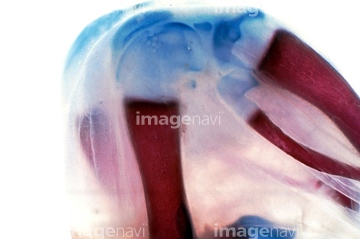
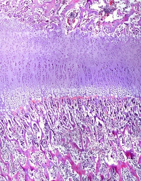
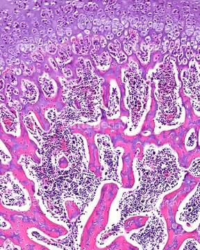
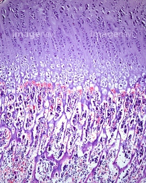
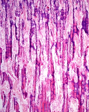
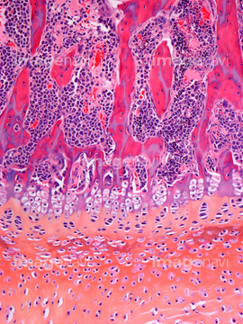
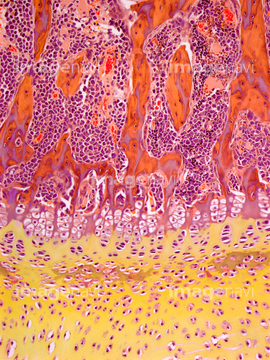
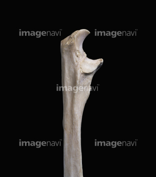
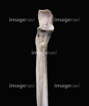
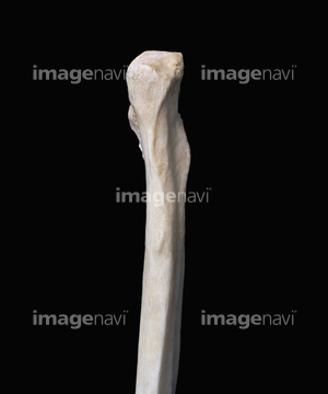
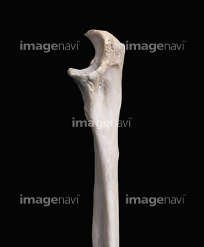
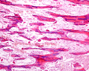
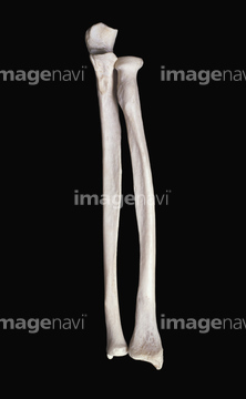
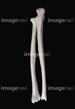
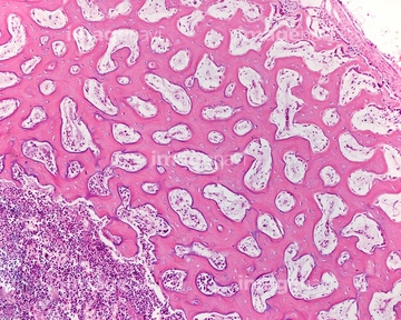
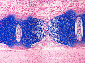
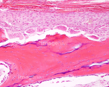
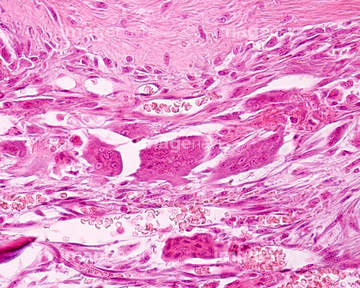
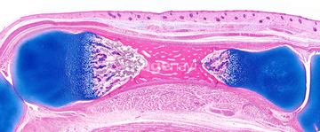
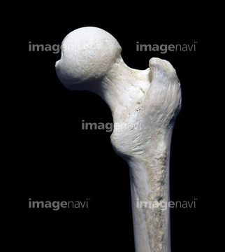
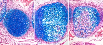
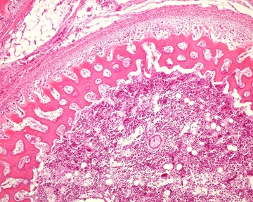
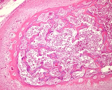
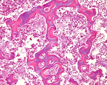
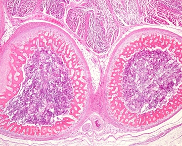
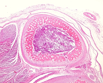
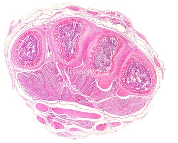
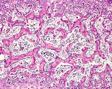
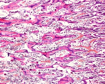
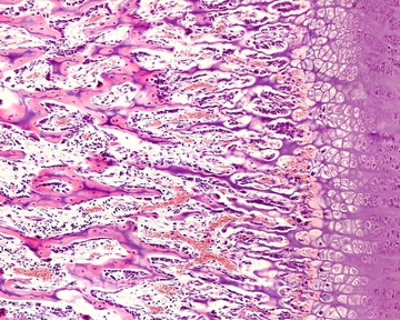
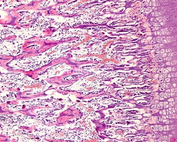
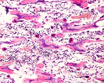
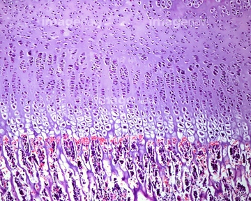
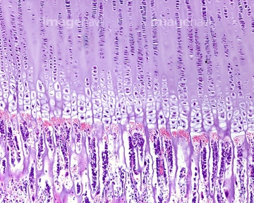
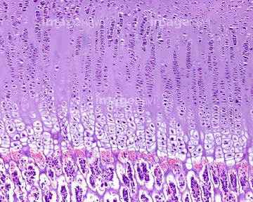
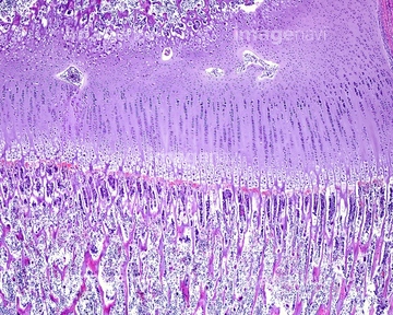
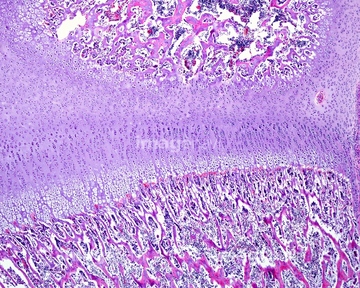
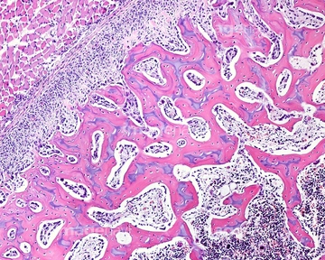
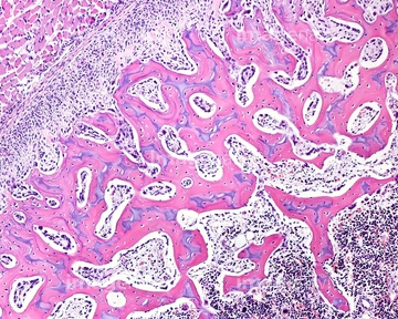
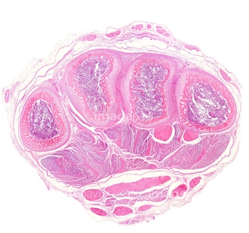
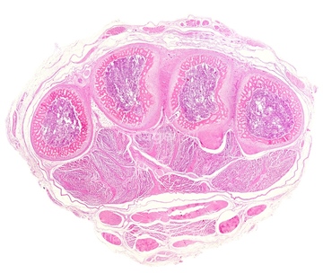
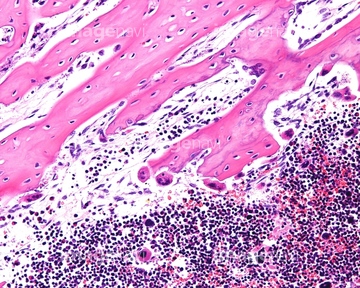
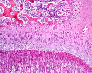
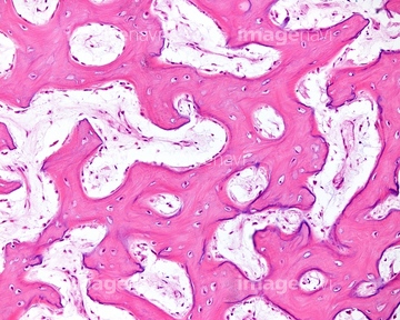
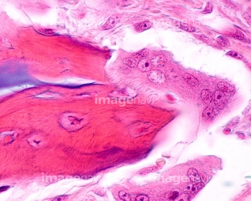
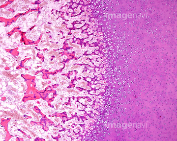
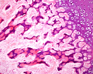
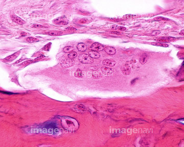
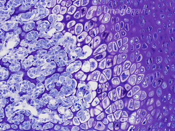
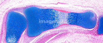
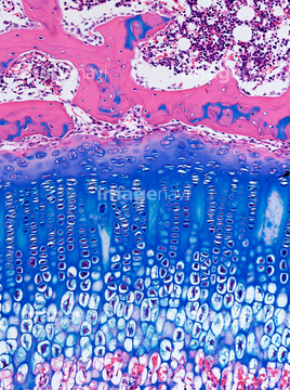
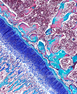
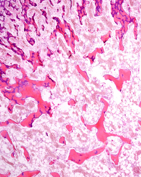
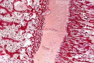
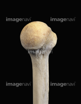
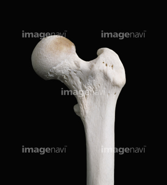
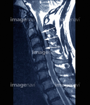
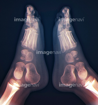
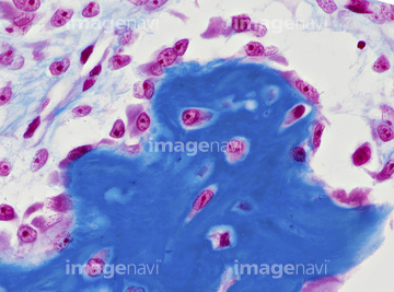
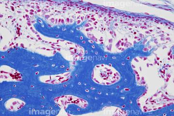
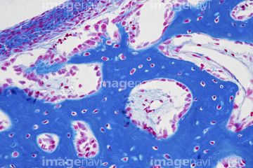
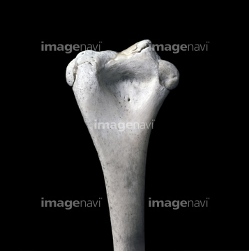
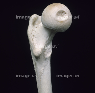
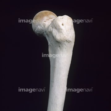
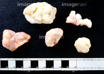
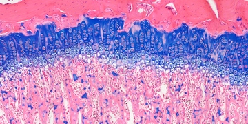
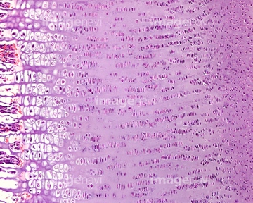
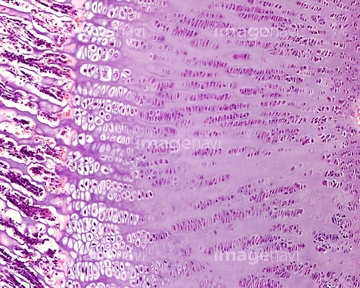
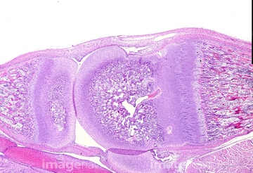
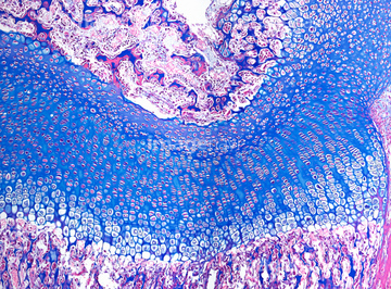
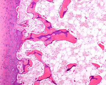
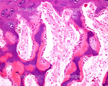
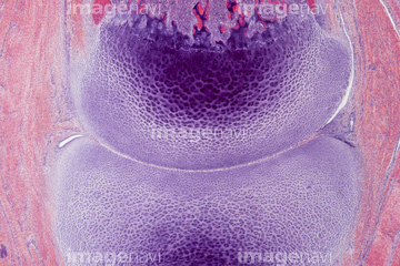
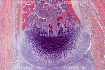
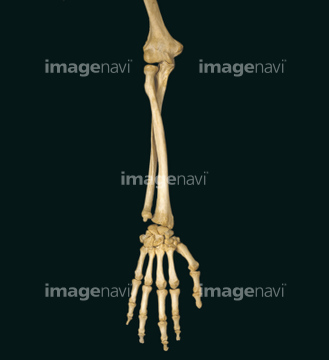
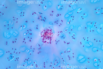
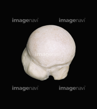
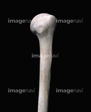
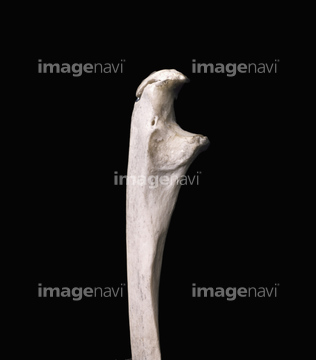
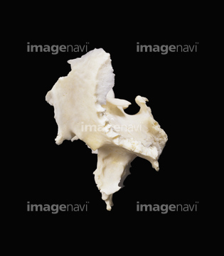
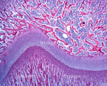
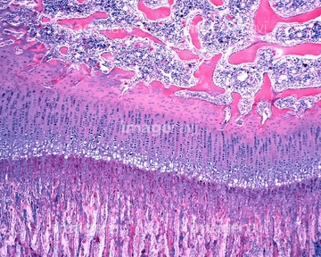
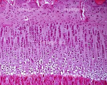
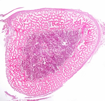
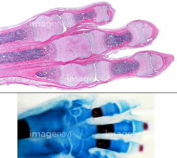
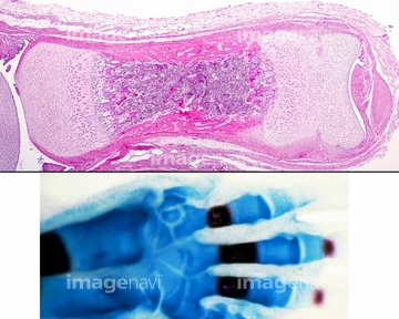
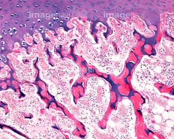
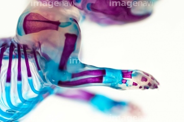
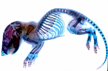
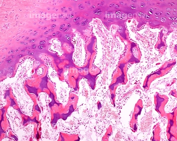
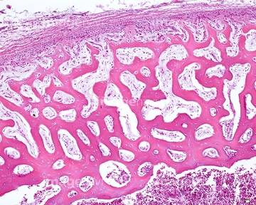
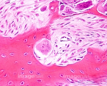
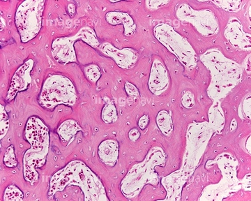
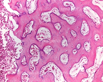
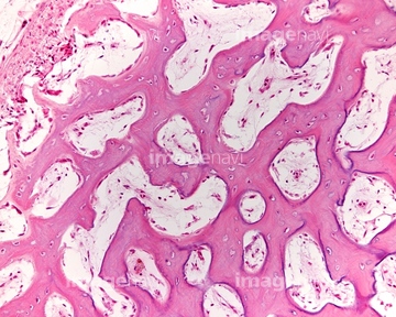
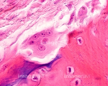
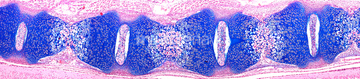
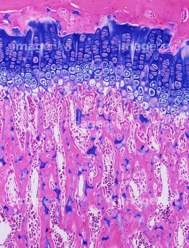
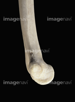
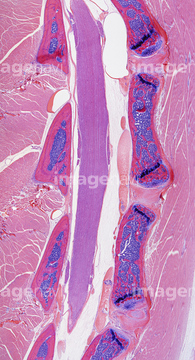
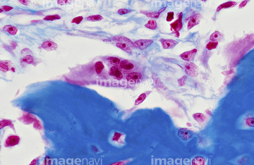
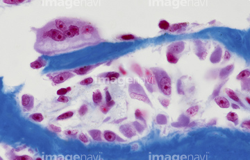
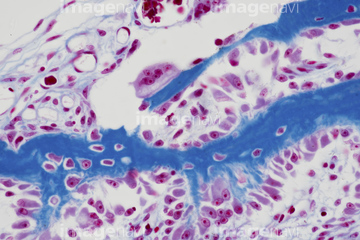
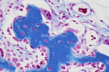
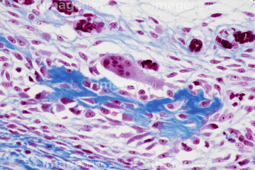
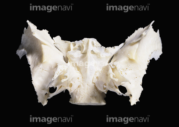
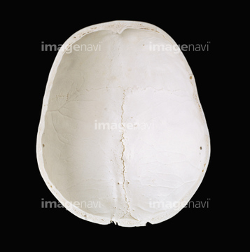
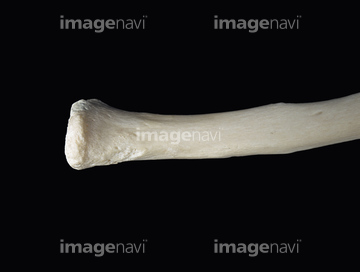
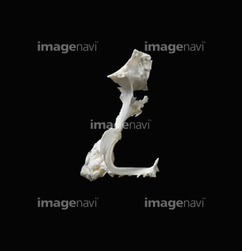
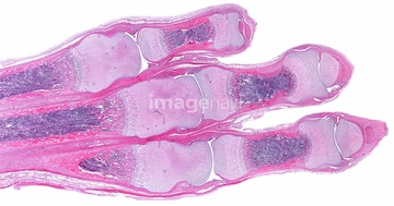
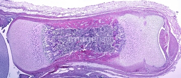
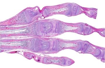
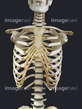
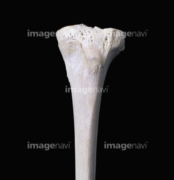
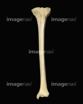
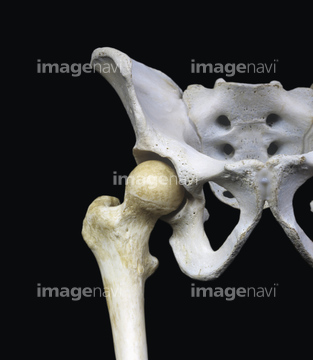
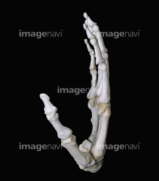
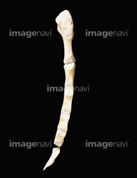
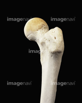
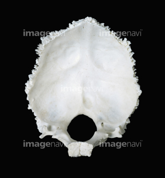
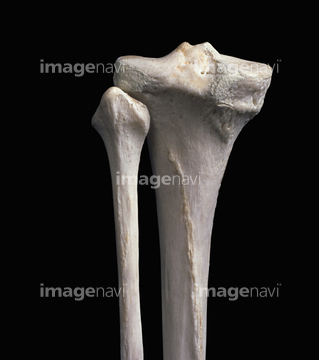
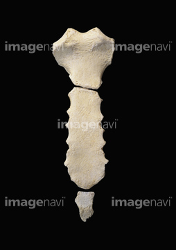
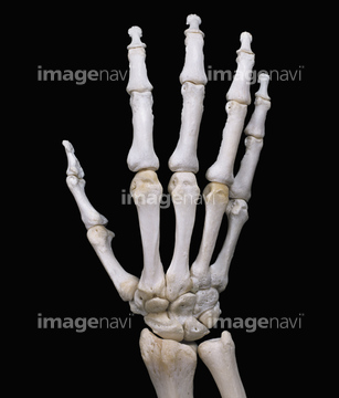
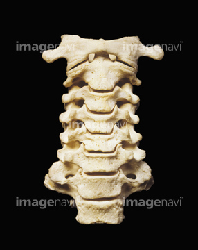
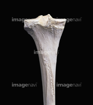
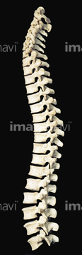
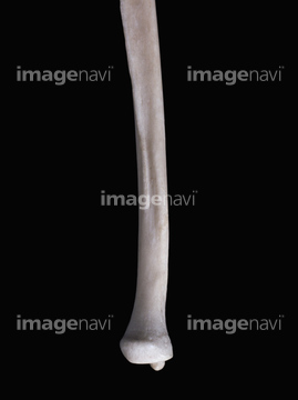
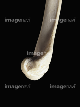
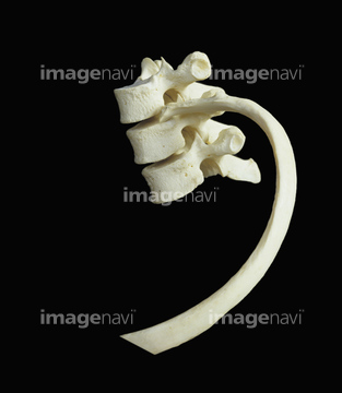
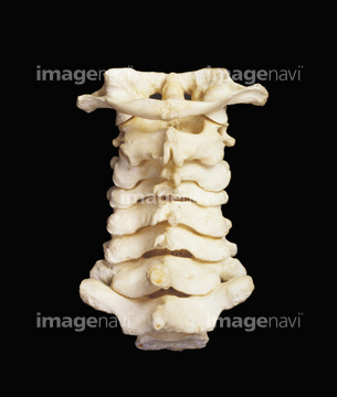
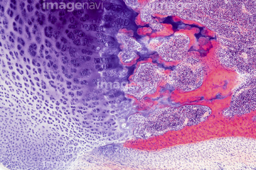
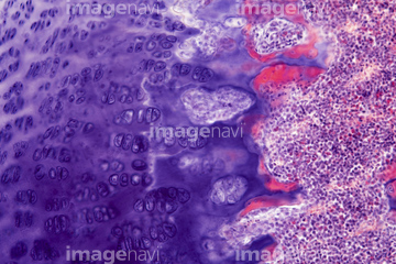
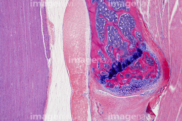
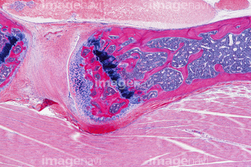
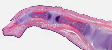
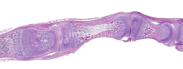
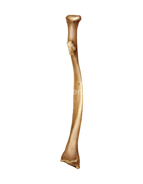
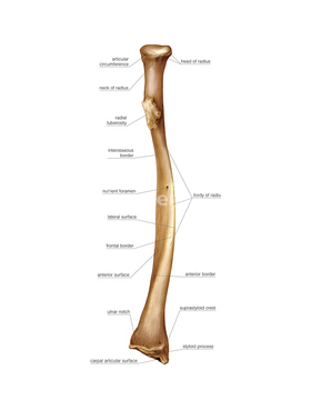
この検索結果には、Articulated right radius and ulna、Embryonic bone diaphysis, light micrograph、Fetal spine, light micrograph、Osteoclasts, light micrograph、Growing bone, light micrograph、Right femur proximal end from behindなどが含まれています。
64225299
64225289
64225367
64225400
64225437
64225395
64225360
64225051
64225050
64060050
64060051
64060052
64060053
64071944
64206764
64206765
64206766
64206767
64225355
64225387
64225419
64225465
64050594

64124003

64116664
64090440
64220946
64093981
64093998
64225329
64225330
64225331
64225332
64225441
64225442

64090452
64260259
64260260
64250018
64225468
64106061


64206787
64206788
64216420
64220958
64161266
64260011
64260251
64260252
64260254
64260257
64251230
64098232
64113106
64225301
64225369
64064521
64235012
64235015
64235016
64225352
64225391
64225397
64013797
64106075
64090154
64090163
64090438
64260253
64225290
64225291
64225353
64225358
64146209
64146210
64146211

64146212
64146213
64146218
64146219
64146220

64220952
64220957
64116641
64225356
64235149
64235017
64235018
64235019
64235020
64235021
64225415
64225440
64225300
64225413
64146214
64224984
64224985
64224987
64224992
64225354
64225357
64225382
64225420
64225438
64225459
64225464
64204266
64224986
64225029
64225062
64225163
64225267
64225281
64225350
64225405
64225466
64234742
64234743
64235150
64235151
64234501
64070775
64070776
| 次ページ |
