HOME > 写真 > 科学・テクノロジー > 科学
10,000件の写真素材が検索されました。
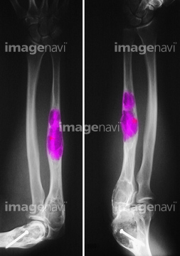
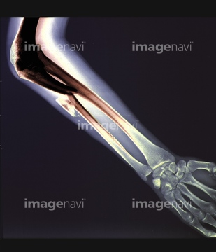
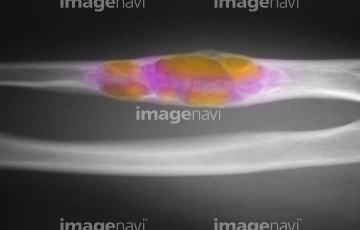
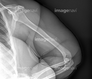
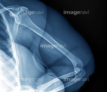
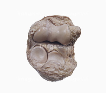
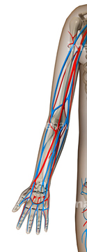
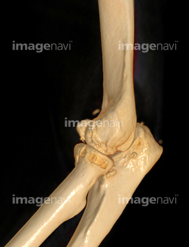
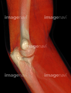
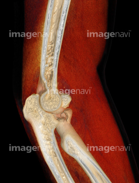
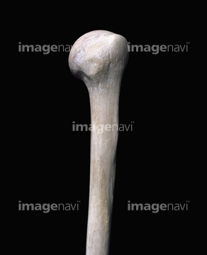
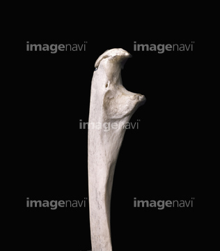
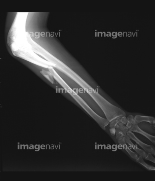
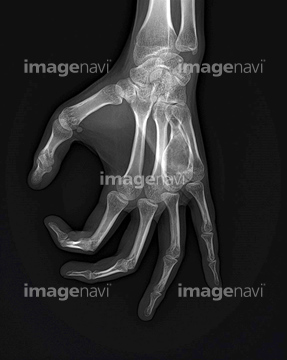
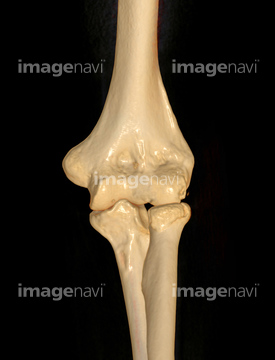
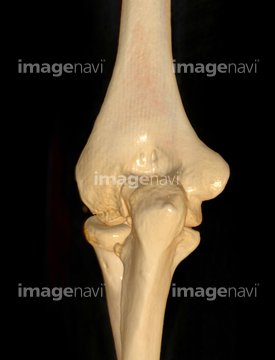
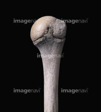
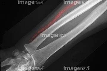
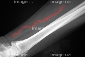
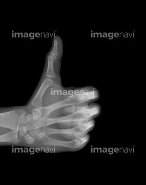
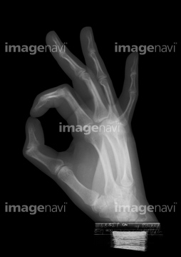
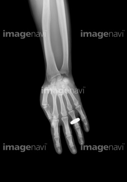
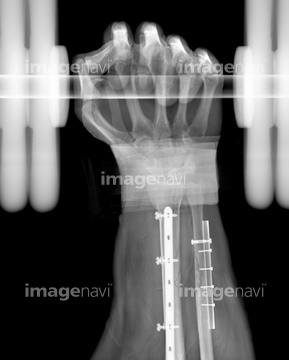
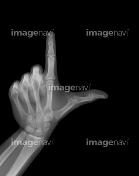
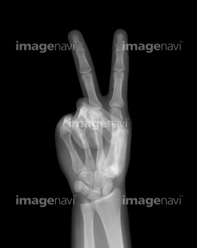
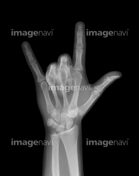
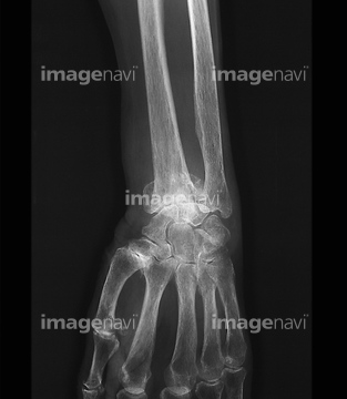
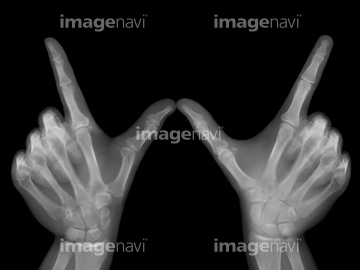
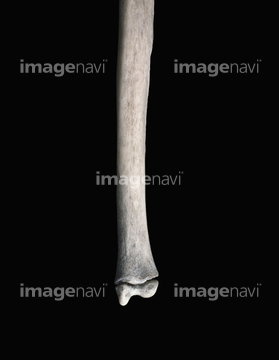
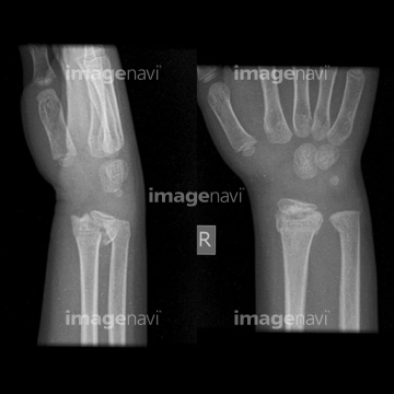
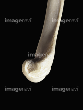
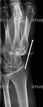
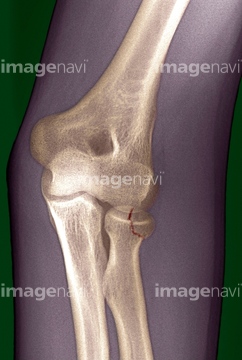
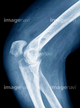
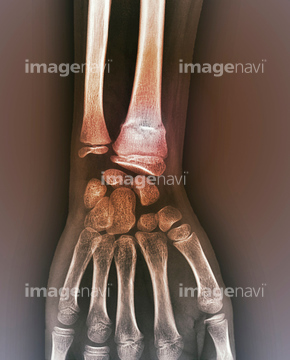
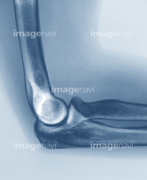
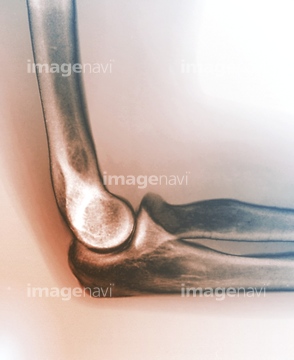
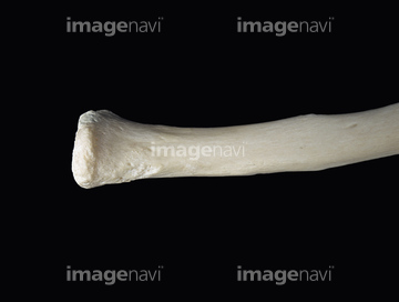
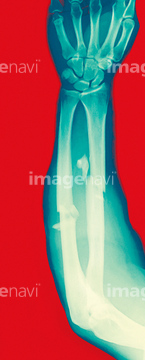
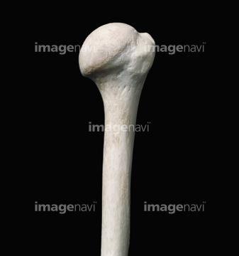
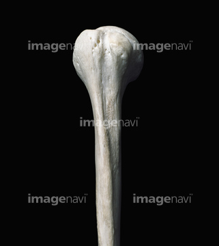
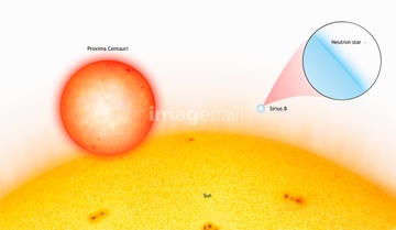
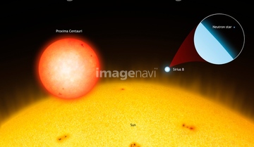
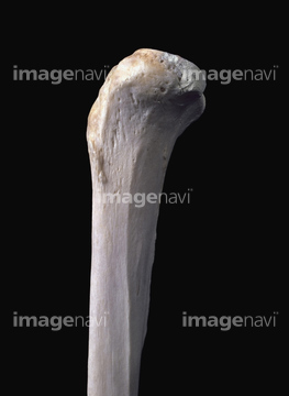
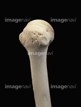
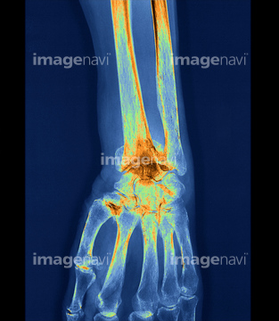
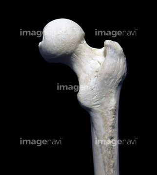
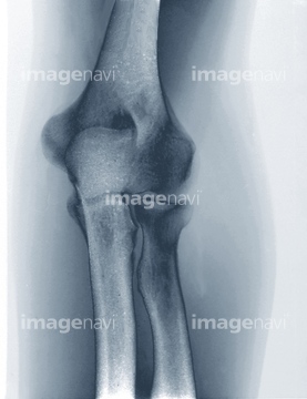
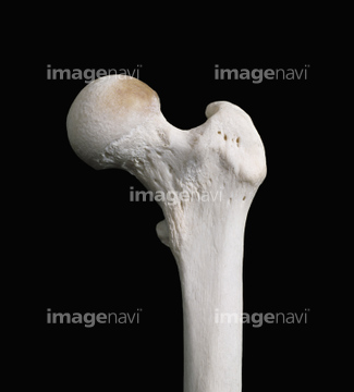
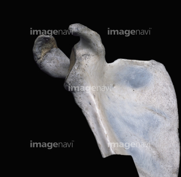
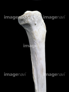
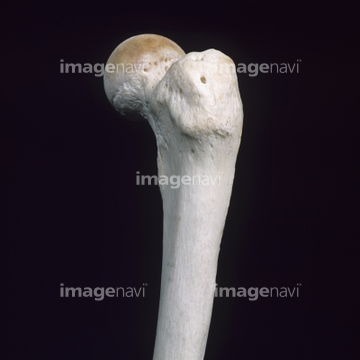
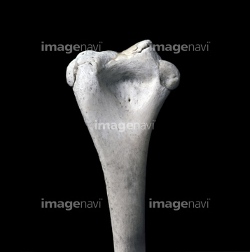
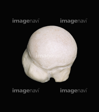
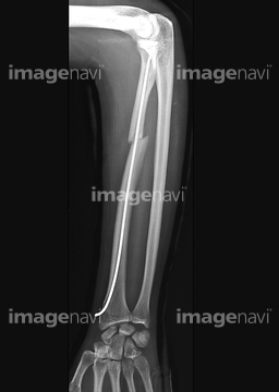
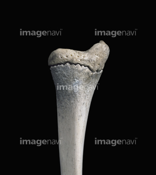
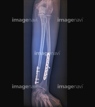
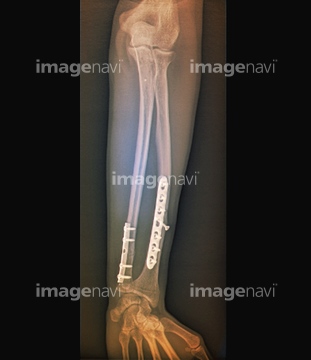
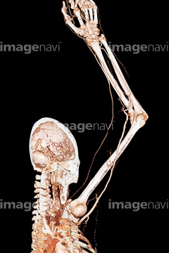
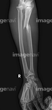
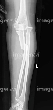
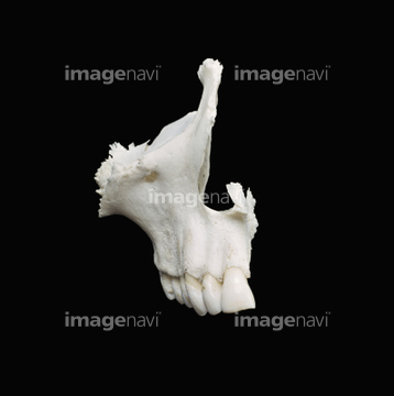
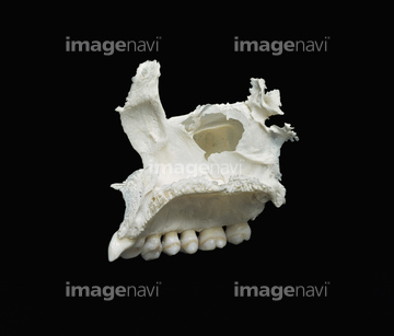
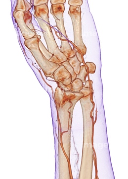
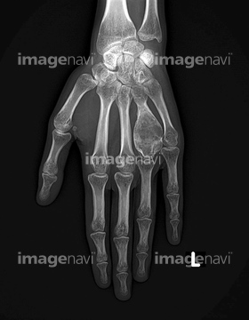
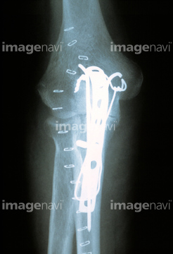
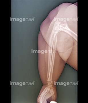
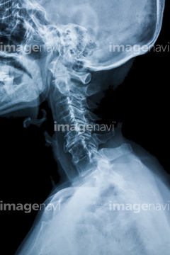
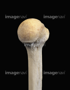
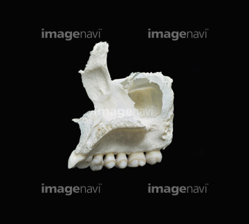
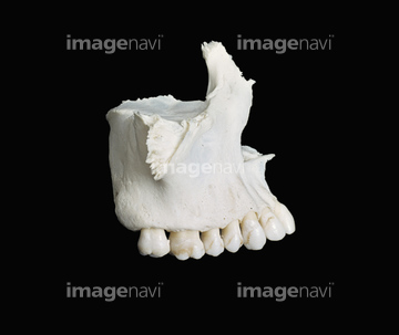
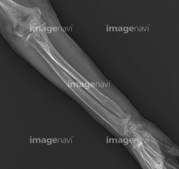
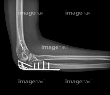
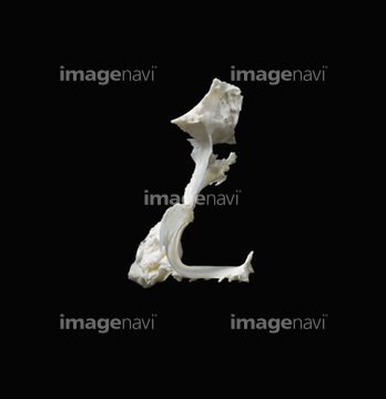
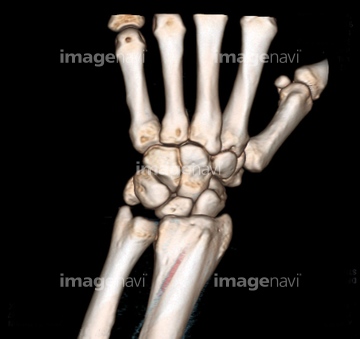
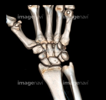
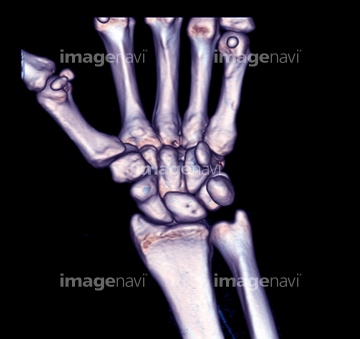
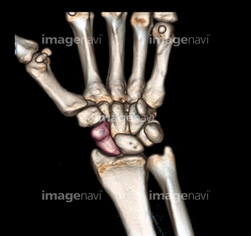
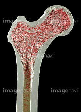
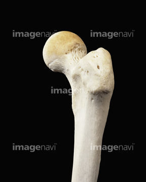
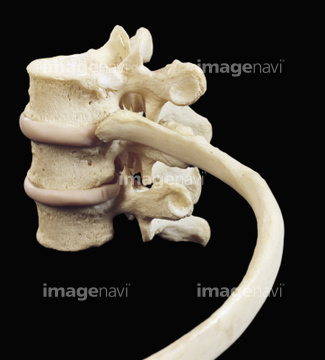
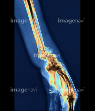
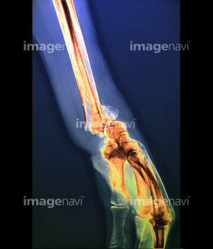
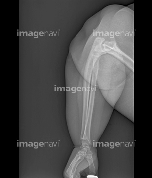
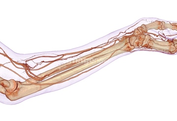
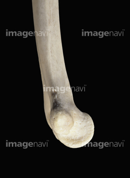
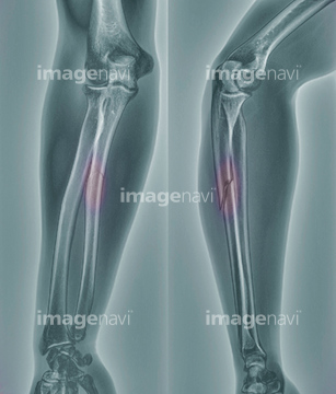
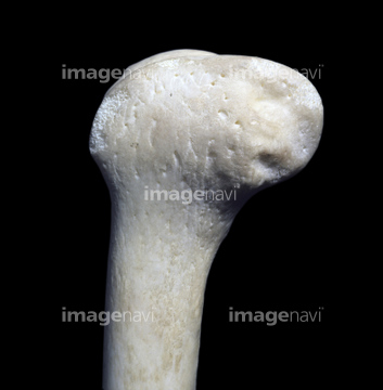
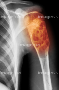
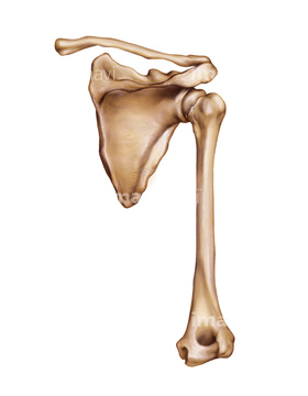
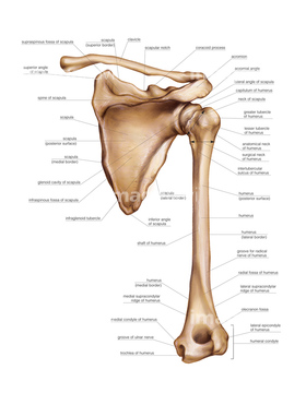
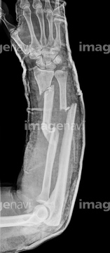
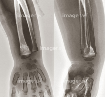
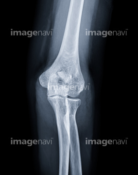
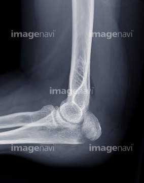
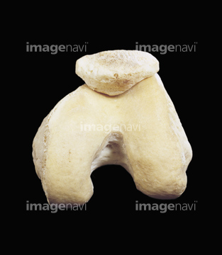
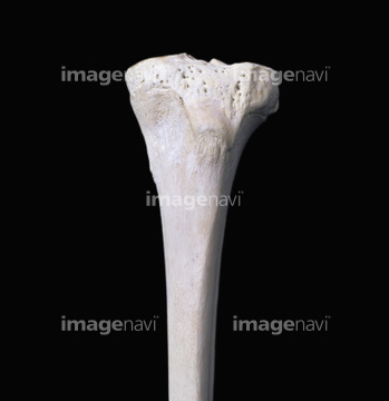
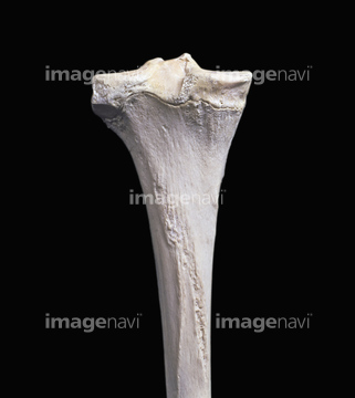
この検索結果には、Healthy elbow joint, CT scan、Humerus proximal right、X-ray, calcific ulnar artery、Thumbs up hands gesture, X-ray、OK hand gesture, X-ray、Hand with jewellery, X-rayなどが含まれています。
64225332
64225329
64225330
64225331
64225442
64225281
64225441
64070769
64070770
64070771
64070772
64070773
64070774
64070767
64070768
64221938
64239259
64228409
64071430

64071433
64070408
64070409
64070438
64070439
64070470
64070471
64070484
64070485
64070488
64070489

64071428
64071429
64225304
64008699
20573684
20573685
20573686
64225291
64225353
64221936
20573682
20573683
64225367
21528531
21528532
20528920
20529192
20527560
20527699
20528706
20528767
20528816
64245397
20528962
64225400
20562167
64225350
64012967
64121464
64099595
64044892
64145062
64185081
64185082
64185083
64098995
64088416
64225299
64225300
17202244
64225295
64225296
64116450
64116451
64225355
64225387
64245386
64224984
64225419
64225468
20562177
20562178
20522171
64185080
64225369
64225360
64225465
64221937
64126205
64225354
64225391
64225397
64225352
64225290
64188132
64188133
64239077
64225029
64225289
64225358
64225437
20548408
20548409
64225376
64225403
64153313
64018421
64188177
16932632
64225301
64225463
64151210
64188131
64225413
64185100
64185102
64185103
64185105
17237153
64225466
64225357
64225382
64225395
64225438
64225464
64225163
64245399
64245400
64188176
64225356
64225377
64225383
20571950
20571951
20572339
20572340
17206973
64225467
64224991
64225390
64225406
64126212
64225016
64225469
64084769
64225402
64225415
64225440
20527600
64185101

64185104
64245398
64070753
64070754
64162705
64221962
20572318
20572320
64225007
64224985
64224987
64224992
64225420
64225459
64225416
64225427
64225428
64225051
64204266
64224986
64225062
| 次ページ |
