HOME > 写真 > 科学・テクノロジー > 科学
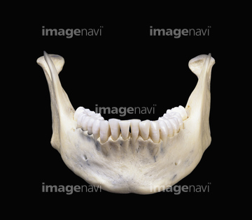
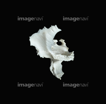
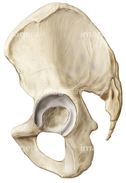
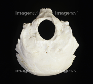
10,000件の写真素材が検索されました。
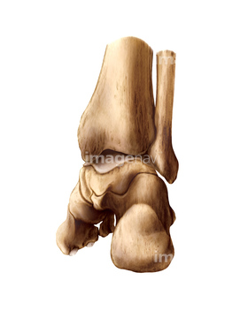
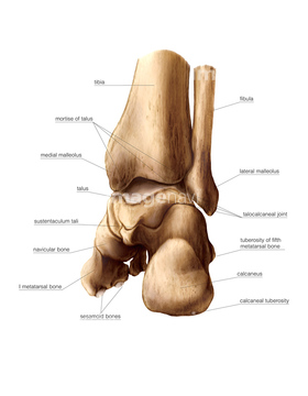
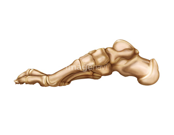
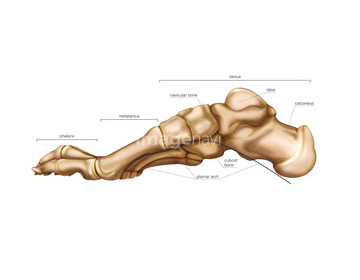
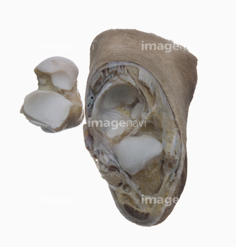
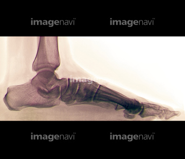
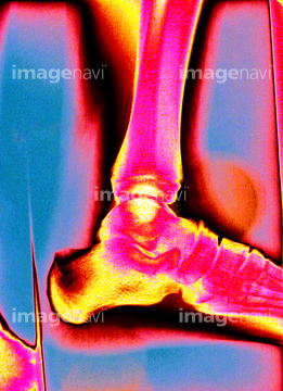
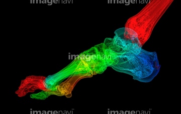
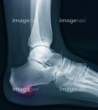
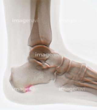
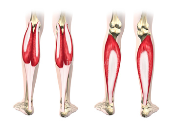
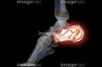
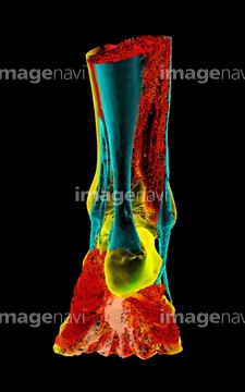
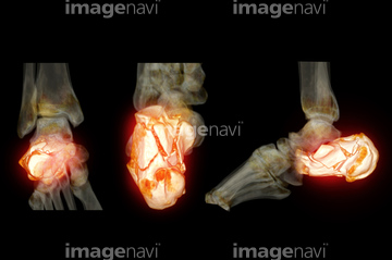
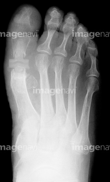
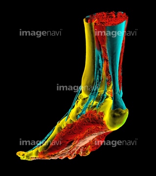
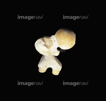
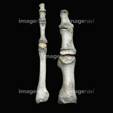
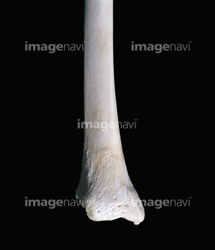
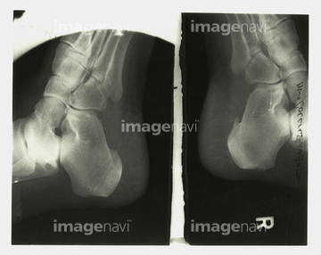
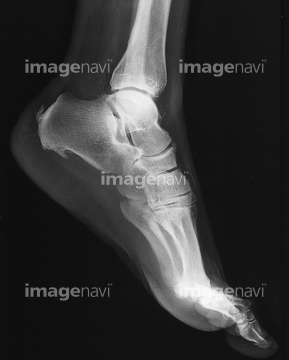
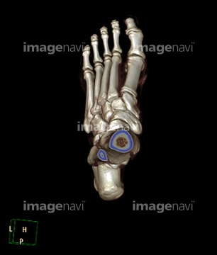
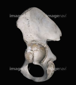
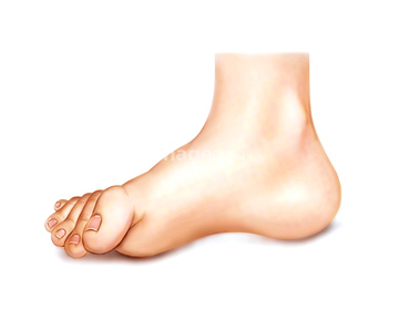
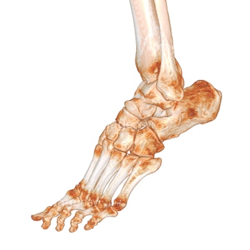
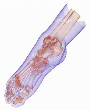
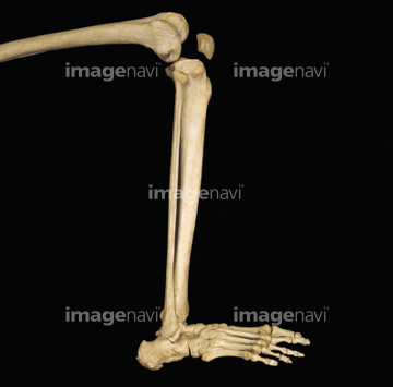
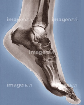
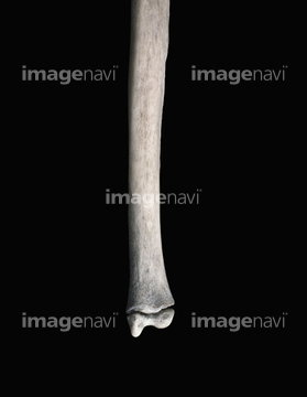
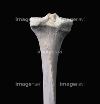
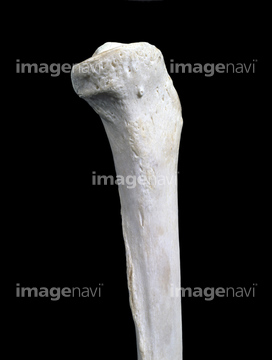
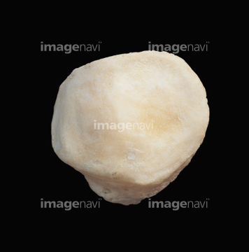
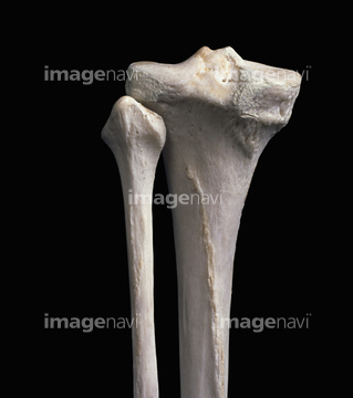
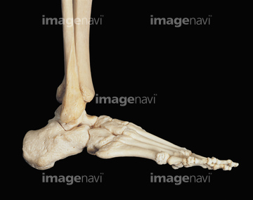
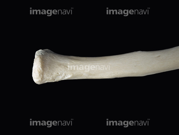
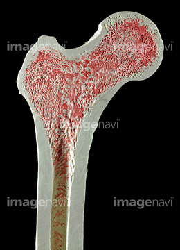
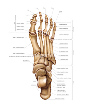
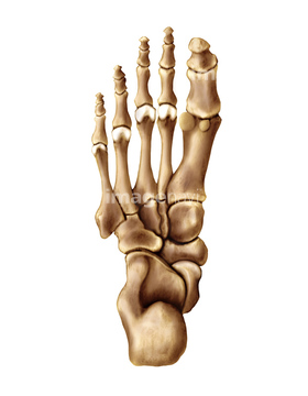
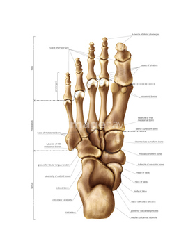
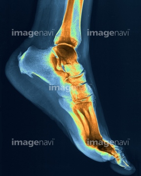
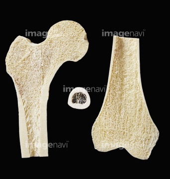
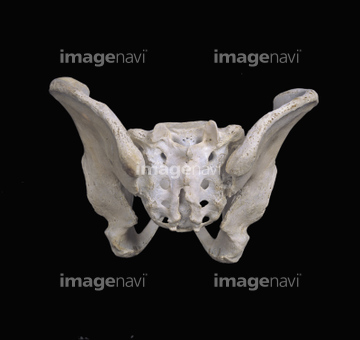
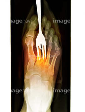
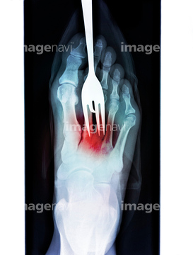
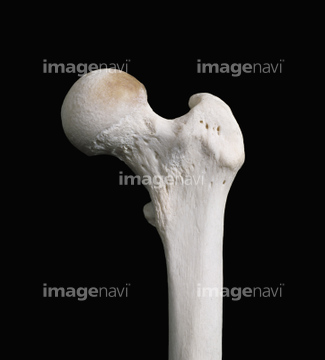
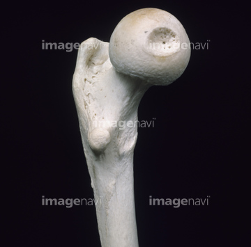
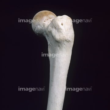
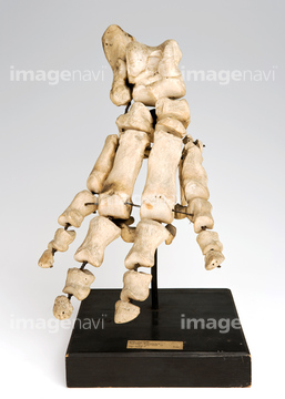
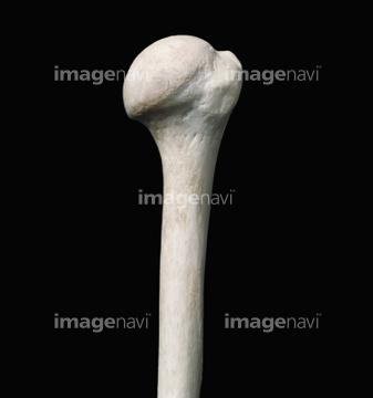
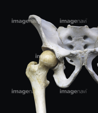
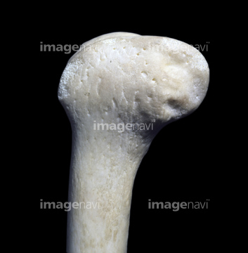
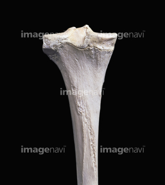
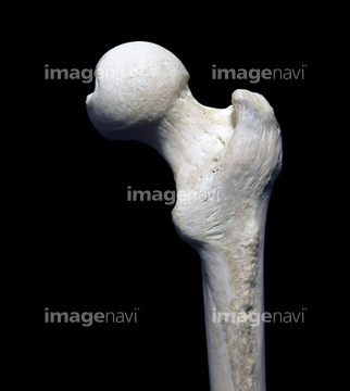
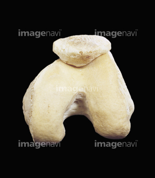
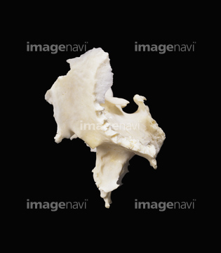
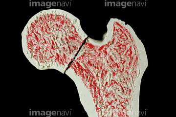
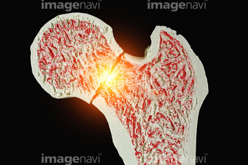
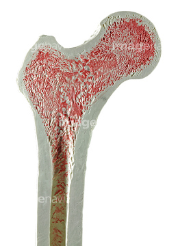
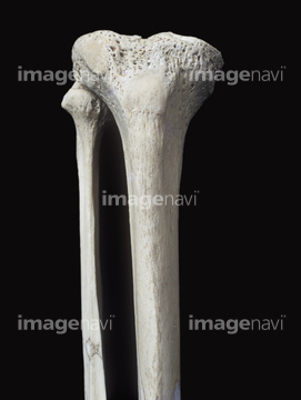
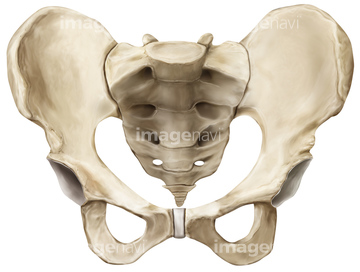
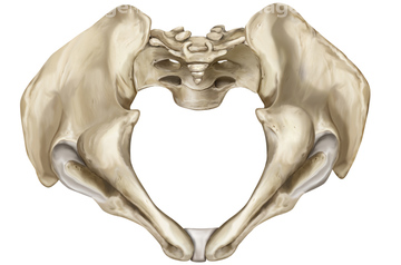
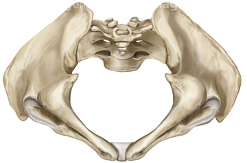
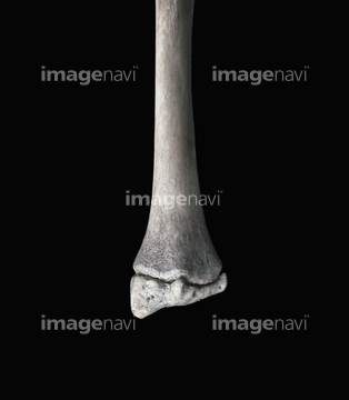
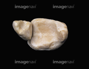
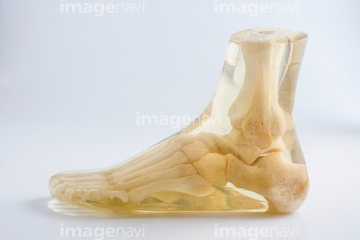
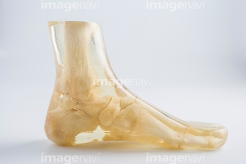
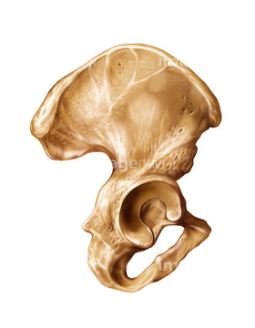
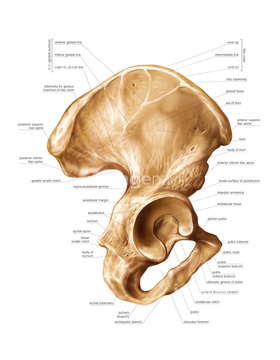
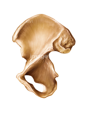
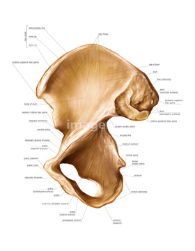
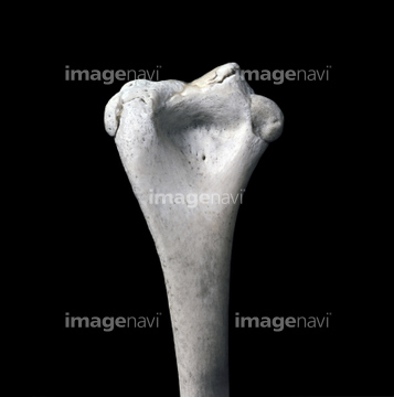
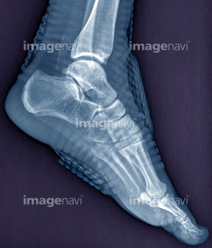
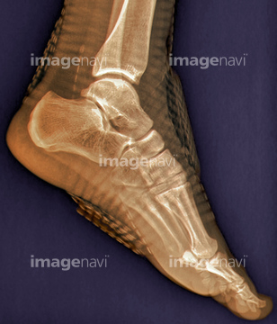
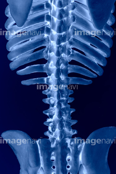
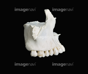
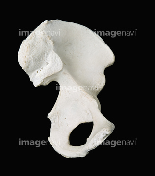
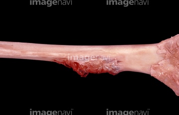
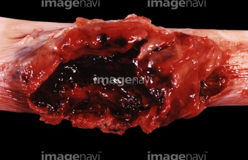
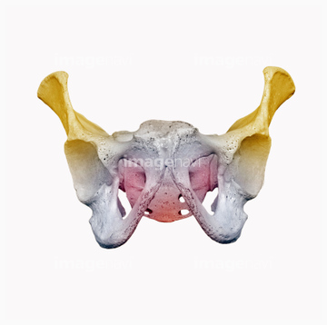
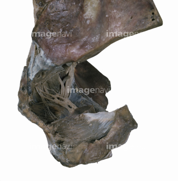
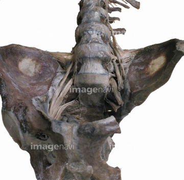
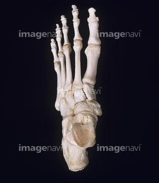
この検索結果には、Lower limb bone, left tibia, from behind、Heel of foot, X-ray、Heel pain, X-ray、Human foot, 3D CT scan、Lateral surface of Hip bone、Bones of the foot, artworkなどが含まれています。
64225390
64225005
64225431
64225364
64070859
64070860
64070853
64070854
64070851
64070852
64070861
64070862
64225393
64049836
64147197
64147198
64147199
64101145
64044851
64044852
64071949
20573645
64044850
64040256
64054597
64168007
20573649
64013806
64056862
64059842
64059843
64059877
64056849
64165610
20527700
64221665

64225297
64225298
20573646
20573647
64165620
64165621
64225392
64225294
64056740
64224125
64221662
64165611
64225395
64070847
64149460
64149461
64149462
64149463
64149464
64168006
64224980
64221663
64225400
64085322
64225301
64225382
64225355
64225419
64225465
64225006
64225438
64225004
64225300
17237153
64070848
64070849
64070850
64221664
64047355
64047358
64225369
64225391
64225397
64093376
64225295
64225296
64224985
64224987
64224992
64225469
64224986
64225468
64225007
64225358
17237145
17237155
20555784
64225271
64225426
64136599

64225003
64224999
64225008
64225009
64225061
17237144
17237158
64225117
64225455
64225415
17237147
64225275
64174369
64174370
64174371
64204257
64225305
53122280
53122748
53122749
64225289
64225291
64225353
64225367
64225437
64225430
64108471
64108473
64225062
64150832
64225285
64225272
64225277
64225461
17237149
64070803
64070804
64070805
64070806
64225404
17237154
53122377
64225443
64225352
17218172
17218173
16934066
64225425
64225403
64225406
64225463
64153736
64153737
64225402
64225408
64225435
64227098
64225424
64225474
53122259
64225292
53156791
53156792
53156793
53156794
17237156
64225118
16934060
64219700
64219701
64219702
64219703
64219704
64219705
64219706
64225432
64225439
64225373
| 次ページ |
