HOME > 写真 > 科学・テクノロジー > 科学
10,000件の写真素材が検索されました。
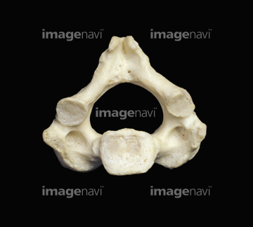
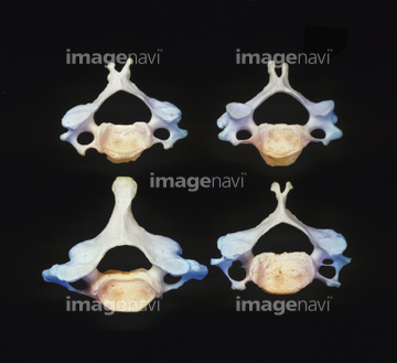
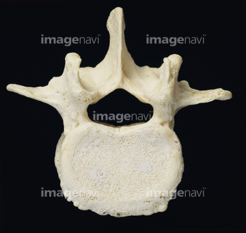
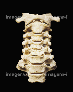
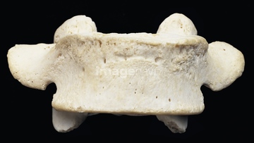
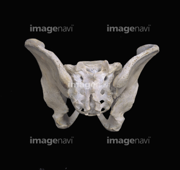
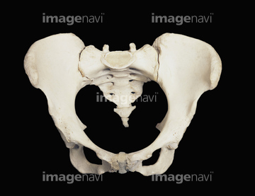
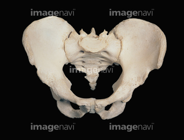
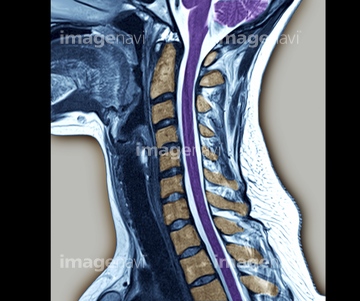
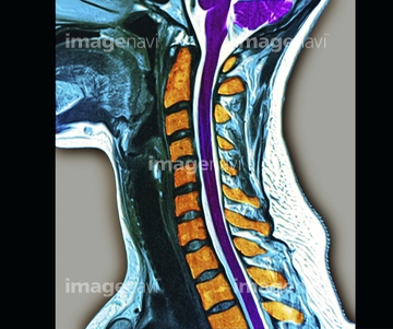
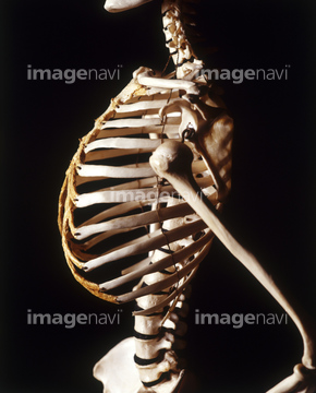
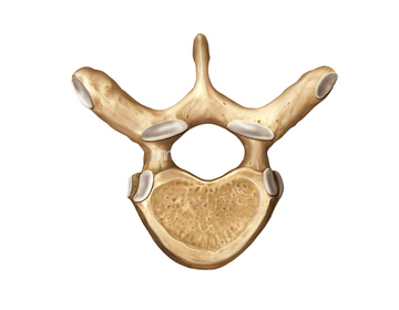
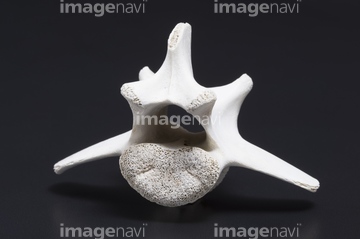
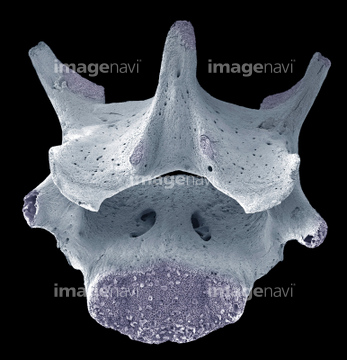
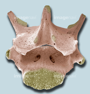
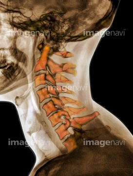
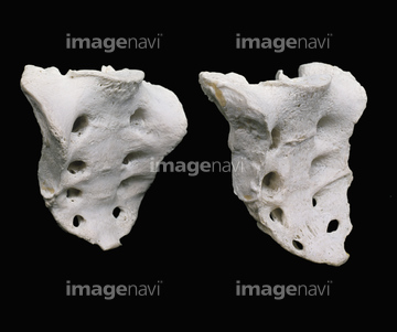
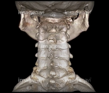
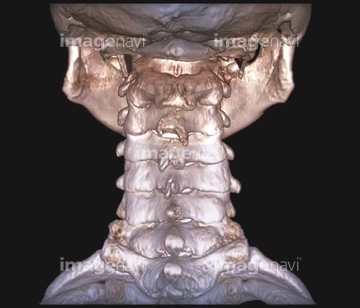
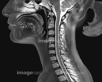
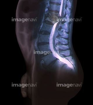
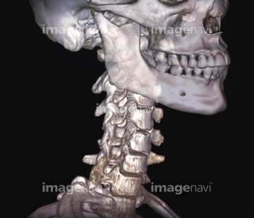
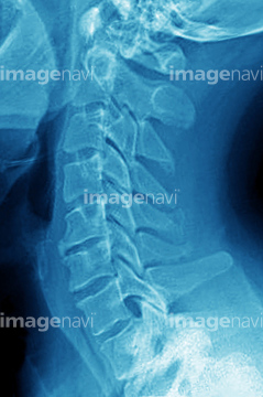
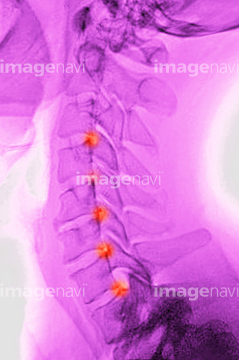
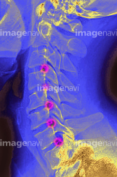
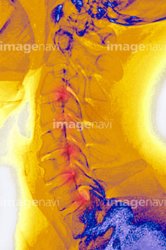
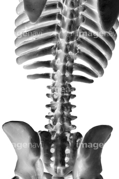
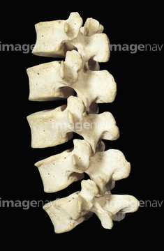
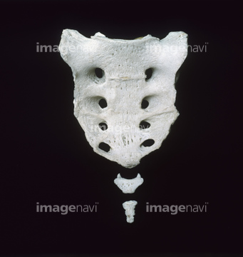
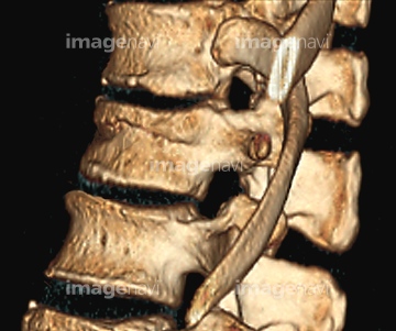
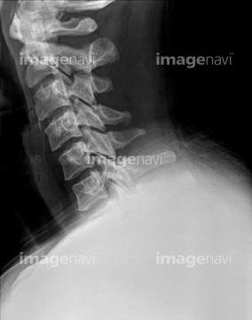
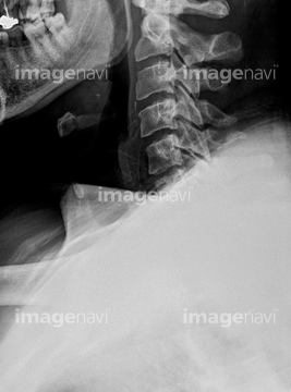
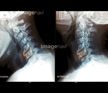
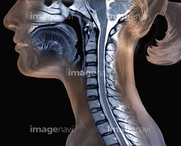
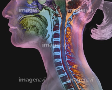
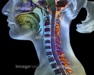
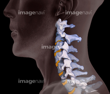
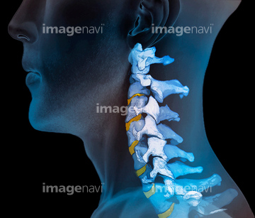
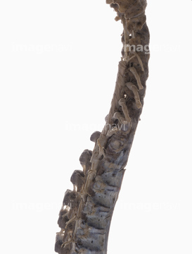
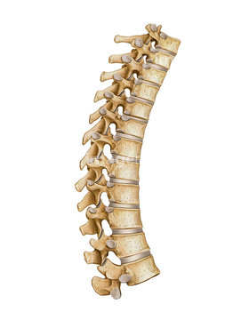
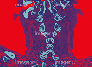
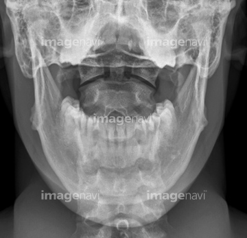
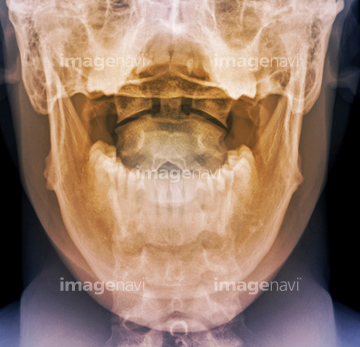
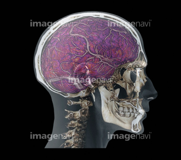
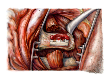
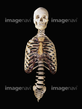
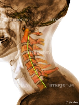
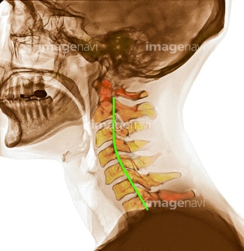
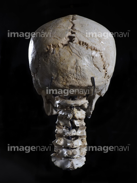
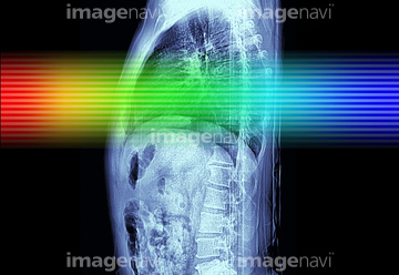
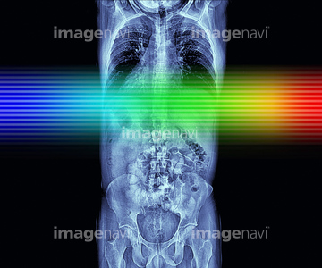
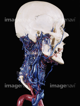
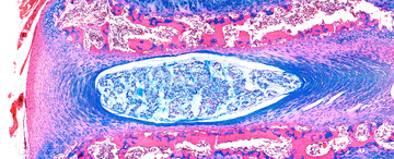
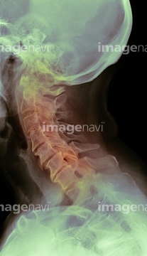
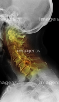
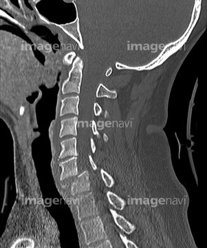
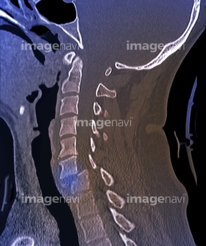
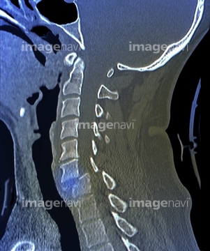
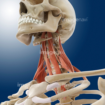
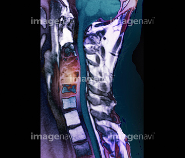
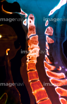
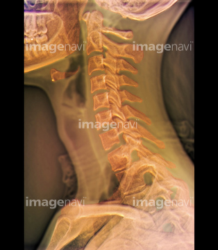
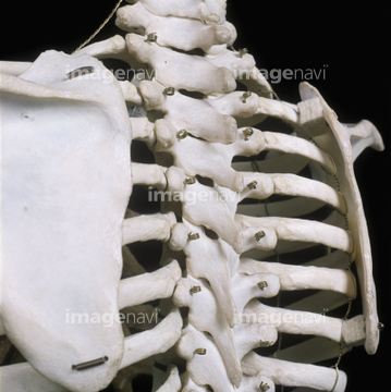
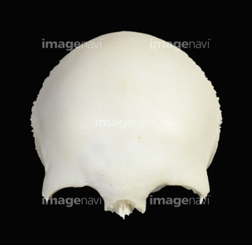
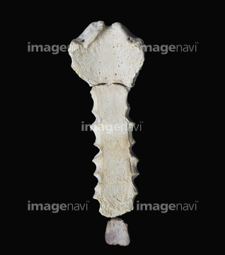
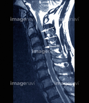
この検索結果には、Malignant tumor localized on cervical vertebrae.、Sacrum、Human cervical spine, 3D CT scan、Cervical spine and brainstem, MRI scan、Pott's disease, MRI scan、Human cervical spine and skull, 3D CT scanなどが含まれています。
64225423
64225386
64225306
64225030
64225031
64225067
64225214
64225163
64225252
64119246
64225253
64225254
64225255
64225008
64225009
64224984
64225456
64225305
64227098
64126417
64126418
64239077
64070715
64070716
64070719
64070720
64070721
64070722
64070723
64070724
64126416
30094706

16934066
64225443
64220883
17202501
17202502
64225271
64165436
64165437
64071441
53157268
53157269
53157270
53157271
16934070
64225027
64070701
64070702
64070703
64070704
64070705
64070706
64070707
64070708
64070711
64070712
64070713
64070714
64264386
64264387
64048259

64126421
64220889
64221654
64221655
64083694
64225408
64225435
64220139
64083701
64083724
64126329
64093994
64130666
17218158
17218159
64225349
64070709
64070710
64054595
64075112
64059844
64059846
64059875
64059876
64071944
20530971
17201767
17205378
17205379
64071698
64072634
64072635
64219678
64077200
64226285
64225406
64225407
64083710
64116638
64090452
64106086
64251241
64165303
64165304
64165305
19907073
64241911
64105764

64075111
64012547
64059845
64064525
17200390
17205370
17205371
17205372
17205380
17205381
64054596
64058535
64058536
64058537
64058539
64040060
64225010
64225011
64225013
64225020
64225287
64064521
17220804
30381768

20547240
64012546
53157357
53157358
53157359
53157360
53157369
| 次ページ |
