HOME > 写真 > 科学・テクノロジー > 科学
10,000件の写真素材が検索されました。
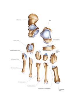
この検索結果には、Bones of the foot, artwork、Broken leg, X-ray、Sacrum and coccyx, pelvic surface、Human foot, 3D CT scan、Lower limb bone, left tibia、Left elbow jointなどが含まれています。
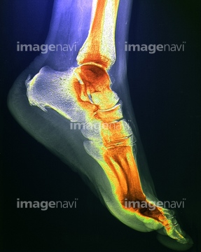
64225431
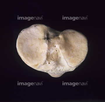
64225364
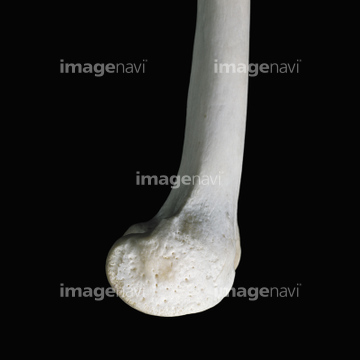
64225393
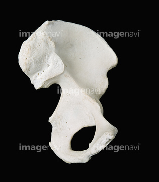
64225390
64225005
64070859
64070860
64070853
64070854
64070851
64070852
64070861
64070862
64147197
64147198
64147199
64040256
64049836
64054597
64225369
64013806
64056862
64071949
64225430
64165620
64101145
64225468
64056849
64059842
64059843
64059877
64044850
64044851
64044852
64168007
64165610
64165611
64225455
64165621
64149461
64149462
64149463
64149464
64168006
64070847
64070848
64070849
64070850
20527700
64225435
64149460
64225392
64225304
64225373
64225374
20573646
20573647
64225391
64225397
64225418
64047355
64047358
64225276
64085322
64225426
64225453
64225356
64225387

64225297

64225308
20573645
64221662
64221665
64225294
64225298
64225402
64225398
64225413
64225355
64225419
20573649
64225465
64225408
64227098
64056740
64224125
64070803
64070804
64070805
64070806
64093376
64225454
64221663
64225271
64225295
64225296
64225382
64224987
64225438
64071953
64225395
64224985
64225372
64225272
64225273
64225274
64225275
64225277
64225416
64225427
64225428
64225452
20521514
64050519
64070807
64070808
64070809
64070810
64070811
64070812
64070813
64070814
64070815
64070816
64070829
64070830
64070831
64070832
64070833
64070834
64070835
64070836
64225445
64070560
64070561
64225006
20521511
20521997
64099597
64221664
17218172
17218173
64101868
64071951
64071952
64071954
64225004
64225287
64060060
64071950
64163385
64225311
64100527
64150078
64070793
64070794
64070795
64070796
64070797
64070798
64070799
64070800
64070817
64070818
64070841
64070842
17237153
64224986
64224992
20521513
64225425
64227767
64106205
30381768
| 次ページ |
