HOME > 写真 > 科学・テクノロジー > 科学
10,000件の写真素材が検索されました。
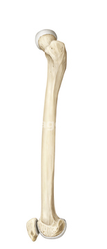
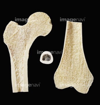
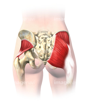
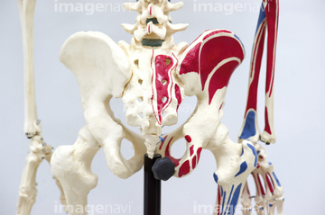
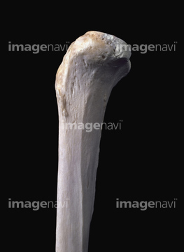
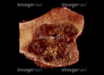
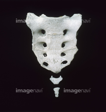
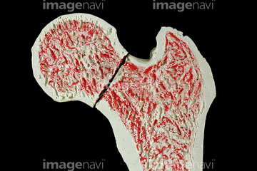
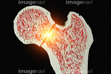
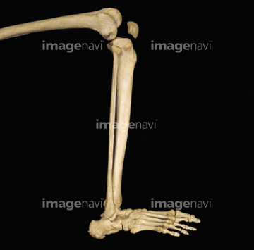
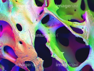
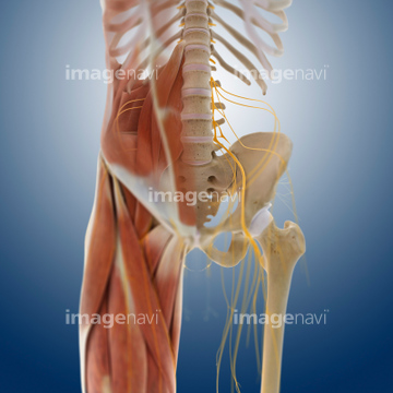
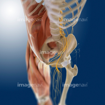
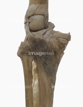
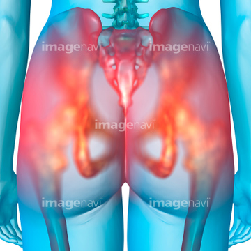
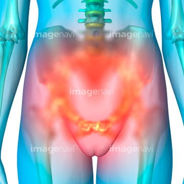
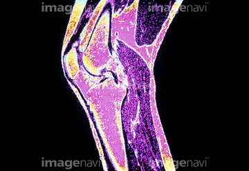
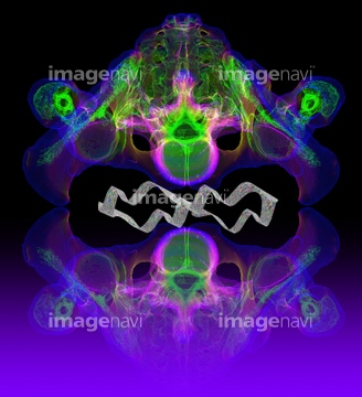
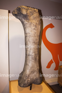
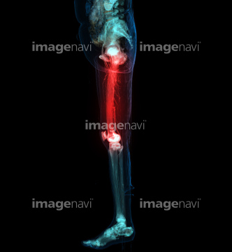
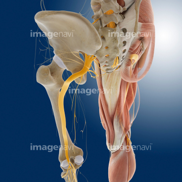
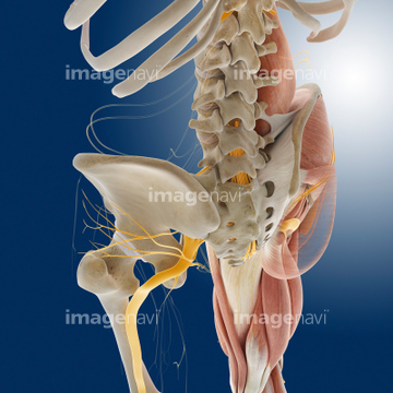
この検索結果には、Fractured hip, artwork、Model of the human knee、4 month old baby's pelvis, X-ray、17 month old baby's pelvis, X-ray、Thigh muscles、Upper right and left femur (leg bone)などが含まれています。
64225468
64225369
64225455
64225391
64225397
64070813
64070814
64225382
64225469
64070807
64070808
64070809
64070810
64070811
64070812
64070815
64070816
64070817
64070818
17237153
64050519
64225298
64225007
64071954
64071951
53122342
64071950
64225419
64225003
64224999
17237147
64040050
64070821
64070822
64074821
64070560
64070561
64070801
64070802
53122649
64100527
32474101
64225465
64060016
64060017
64060018
17237144
17237158
64225117
64078969
64078973
64249964

20530969
64063347
64225355
64225431
64225435
17237145
17237155
64224980
64150078
64060019
64060020
64225370
64224986
64049300
64049302
64056684
64219700
64219701
64219702
64219703
64219704
64219705
64219706
64219707
64219708
64064641
64056686
64056687
64049860
64064639
64064640
64071956
53122377
64225418
64056862
64085233
64236964
64070841
64070842
64064637
64064638
64057264
64057265
64057271
64057848
64057849
64057850
64159940
64056685
20555788
64060015
17237156
64225118
64260899

64225297
64225392
64013810
64013813
64013814
64013815
64071949
64101867
32474104
17237154
64049831
64049847
64159939
64070847
64224985
64150079
64147197
64147198
64147199
64225294
64070848
64070849
64070850
64070861
64070862
64014105
64048561
17237149
64059505
64060021
64070829
64070830
64070831
64070832
64070833
64070834
64070835
64070836
64010066
64071469
64071470
64054597
64225430
64224987
64225413
64225295
64225296
64012562
64246493
64245405
64245406
64012992
64225364
64225408
64227098
64149495
64060267
64060268
64060269
64049832
64070853
64070854
64260893
64044871
64071952
64071953
20569560
64060023
64060024
64060025
64060026
| 次ページ |
