HOME > 写真 > 科学・テクノロジー > 科学
10,000件の写真素材が検索されました。
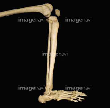
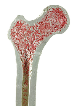
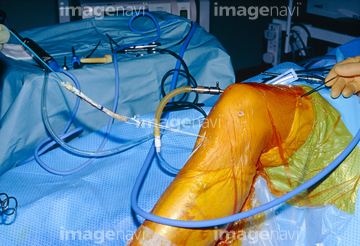
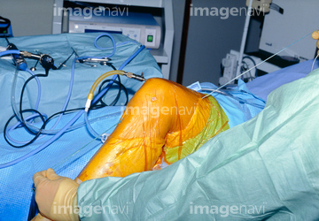
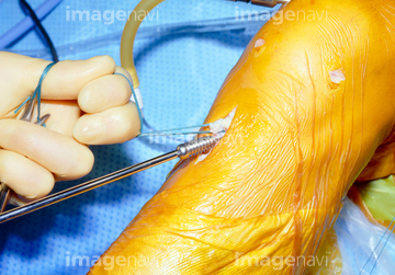
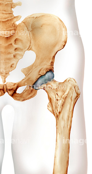
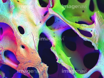
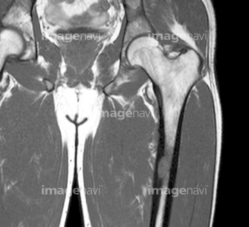
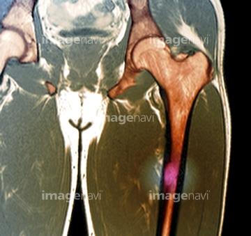
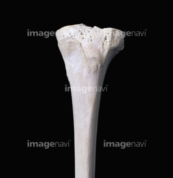
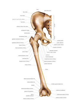
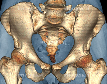
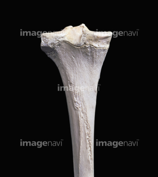
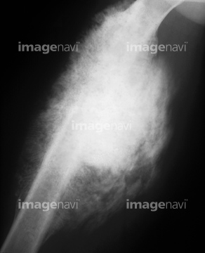
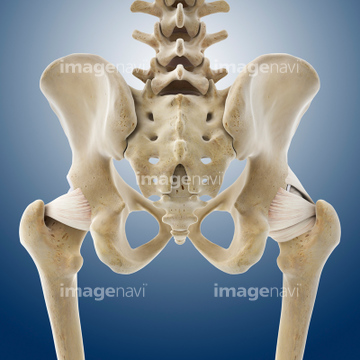
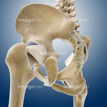
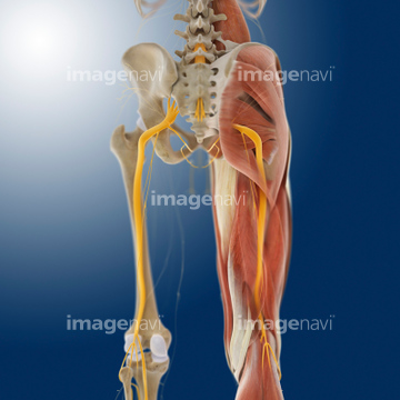
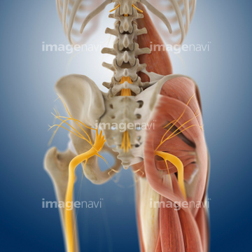
この検索結果には、Hip, 3D bone reconstruction from MRI Data、Knee ligament replacement surgery、Healthy hip, artwork、Lower limb bone、x-ray showing femur fracture from auto accident、Bones of the left footなどが含まれています。
64225369
64225382
64225468
64225391
64225397
64225469
64225298
17237153
64225455
64225007
64224999
64225003

53122342
53122649
64224980
17237147
64225294
64070807
64070808
64070809
64070810
64070811
64070812
64070813
64070814
64070815
64070816
64070817
64070818
64225006
53122377
64225390
64225419
64225392
64225465
64225431
17237144
17237145
17237155
17237158
64225117
64225430
64225355
64013810
64013813
64013814
64013815
64050519

64225297
64225364
64219700
64219701
64219702
64219703
64219704
64219705
64219706
64224987
64225356
64225387
20555788
64070560
64070561
64048561
64014105
17237149
17237156
64225118
64225370
64059505
64071954
64012992
64071950
17237154
64060016
64060017
64060018
64060019
64060020
64225438
64049831
64049832
64049847
64049874
64150078
64224985
64221968
64221969
64221970
64064639
64064640
64071951
64219707
64219708
64070821
64070822
64078969
64078973
64012562
64070801
64070802
64049860
64224986
64044871
64056684
64100527
64056686
64056687

64060022
64060023
64060024
64060025
64060026
64060027
64060028
64225395
64260899
64063347
64064637
64064638
64064641
64010066
64040050
64071469
64071470
64071693
64071695
64071696
64074821
64012607
64012608
64012614
64012615
64049300
64049302
64060015
64060021
64225393

30342249
64109321
64008672
17237157
64246493
64225295
64225296
64249964
64012525
64012526
64012604
64012605
64012606
64012989
64012993
64013012
64014101
64014102
64225008
64225009
20530969
17236828
17236829
32474101
64084815
64084817
20555784
64044869
64044870
64225005
64225301
17218136
17218137
20559313
20559314
20559315
20559316
20559317
20559318
20559319
| 次ページ |
