HOME > 写真 > 科学・テクノロジー > 科学
10,000件の写真素材が検索されました。
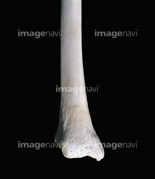
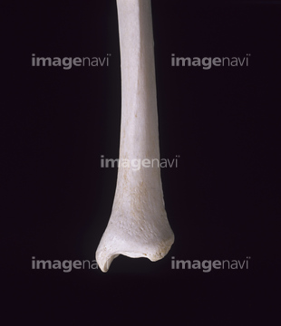
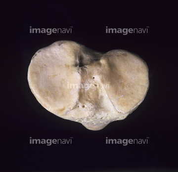
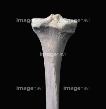
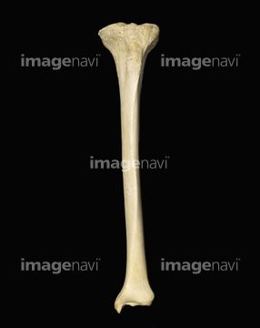
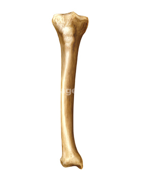
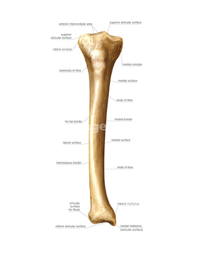
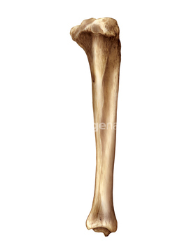
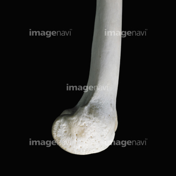
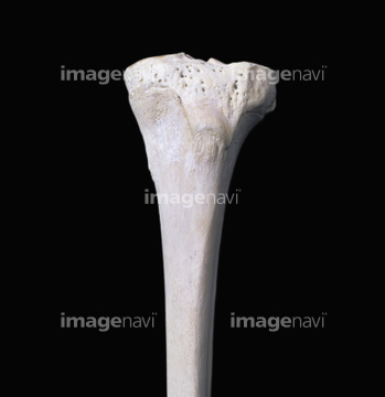
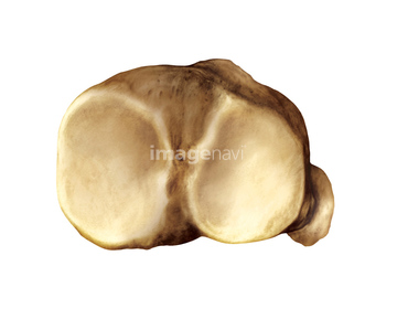
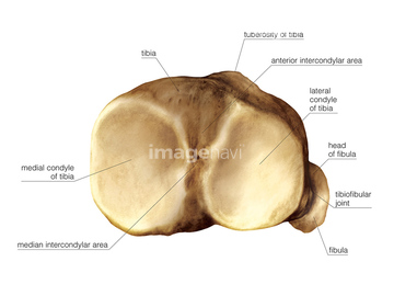
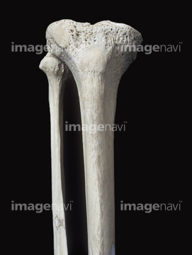
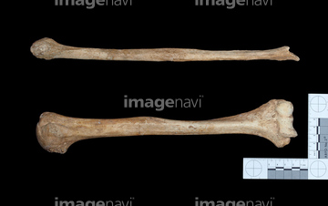
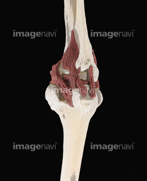
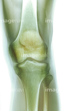
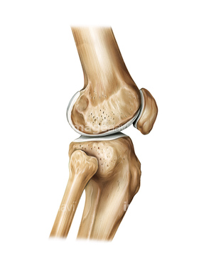
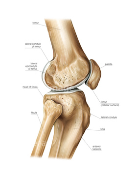
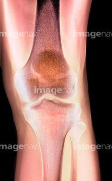
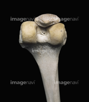
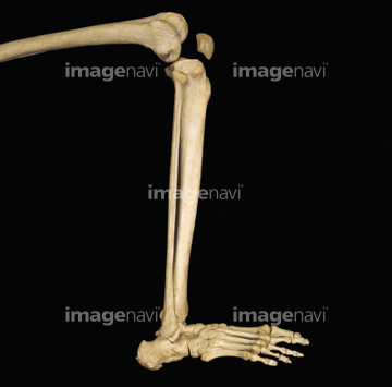
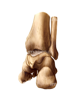
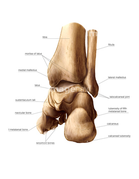
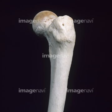
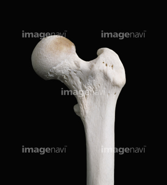
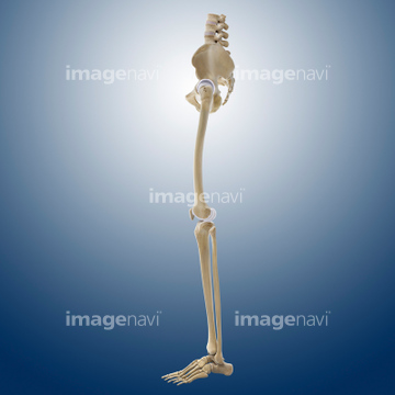
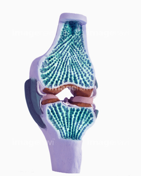
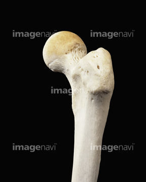
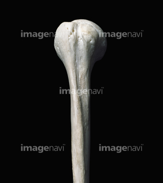
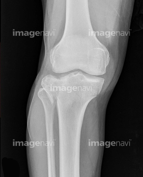
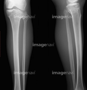
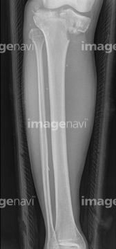
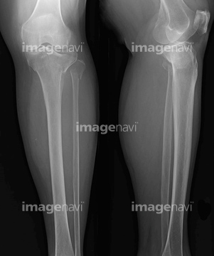
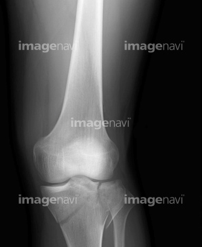
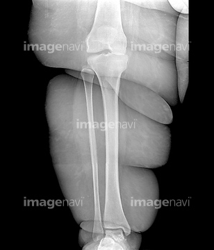
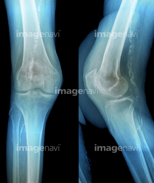
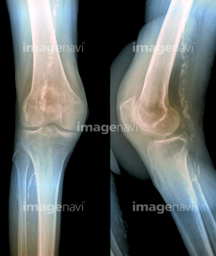
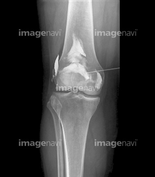
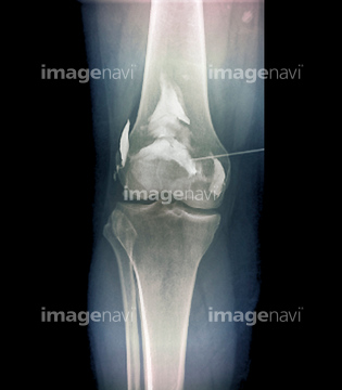
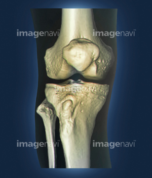
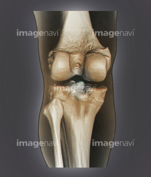
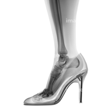
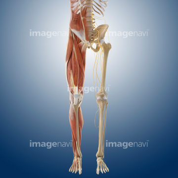
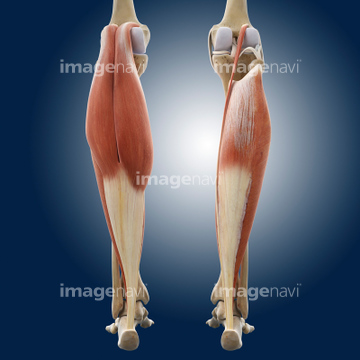
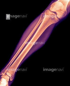
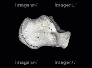
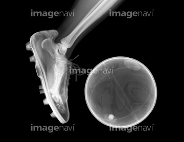
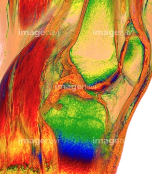
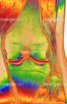
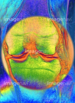
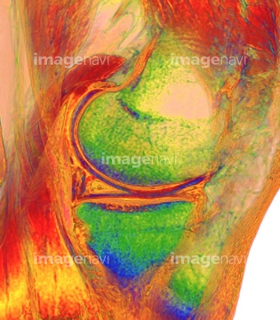
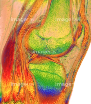
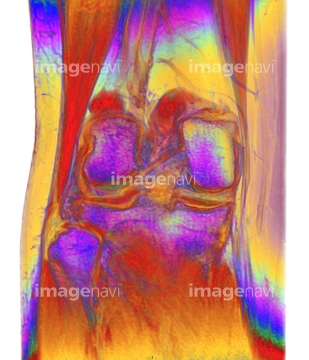
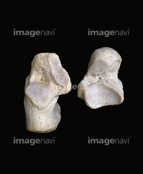
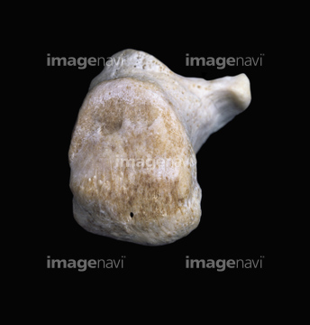
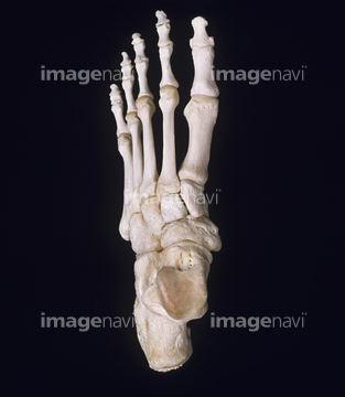
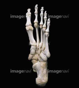
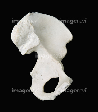
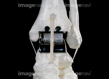
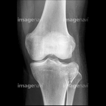
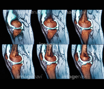
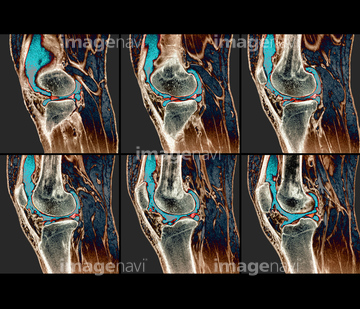
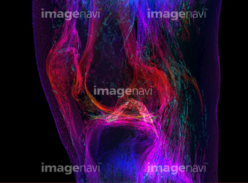
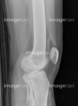
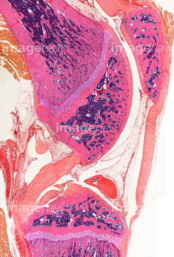
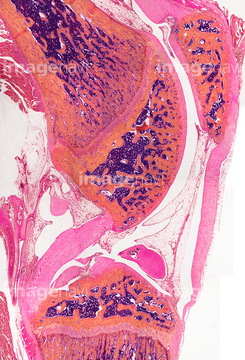
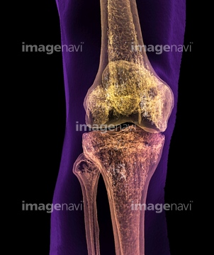
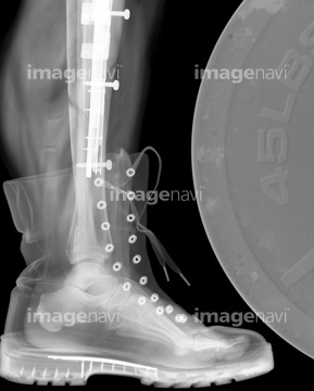
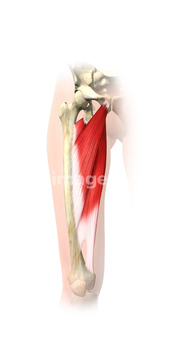
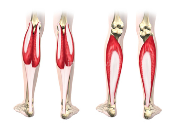
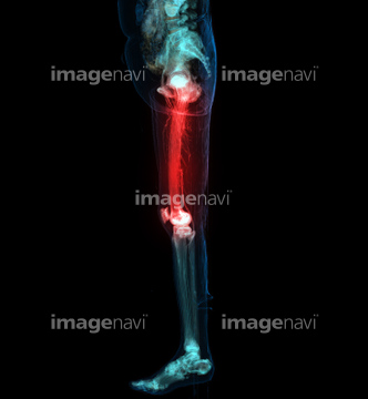
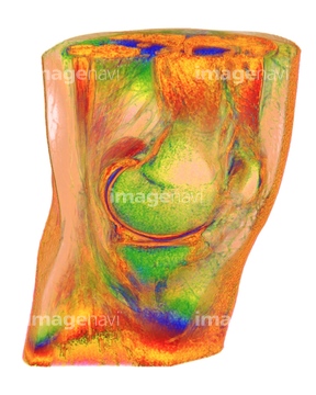
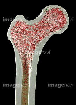
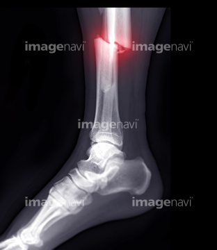
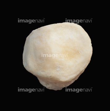
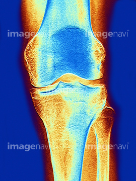
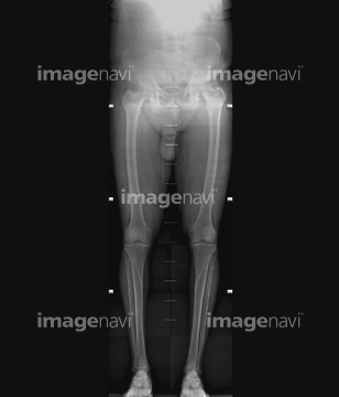
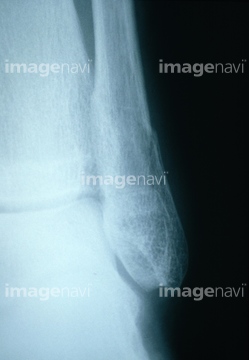
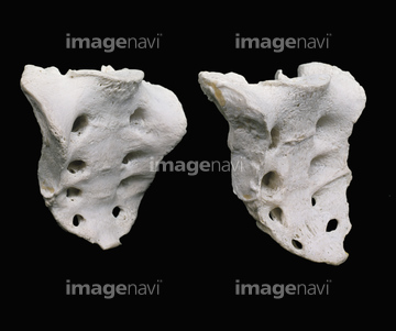
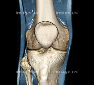
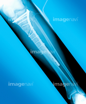
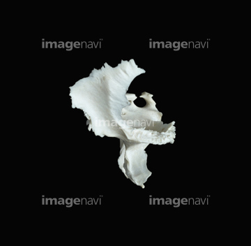
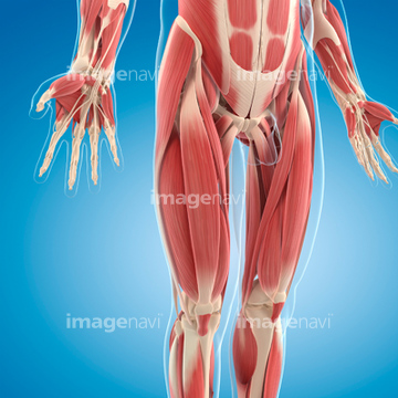
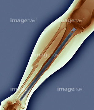
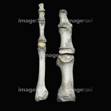
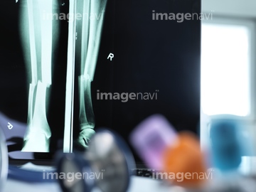
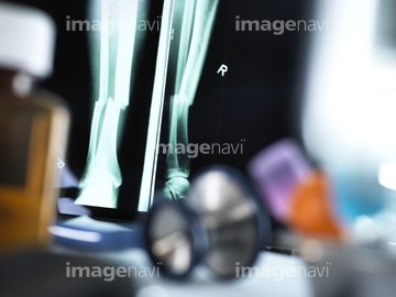
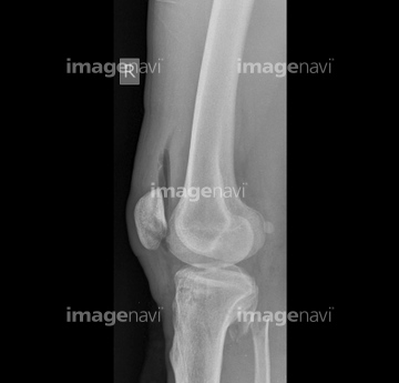
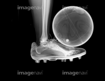
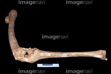
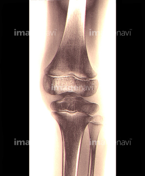
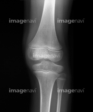
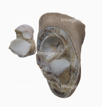
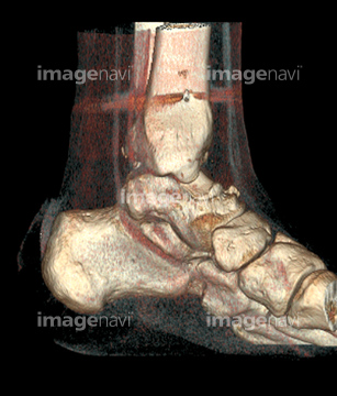
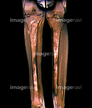
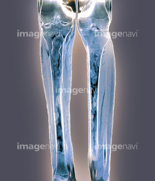
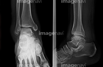
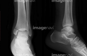
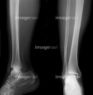
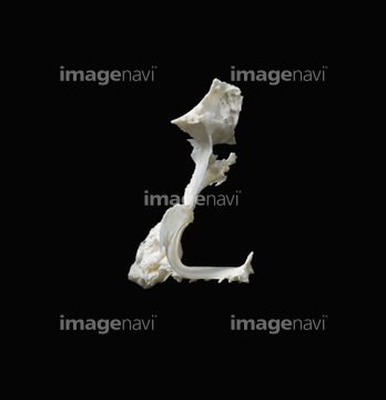
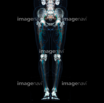
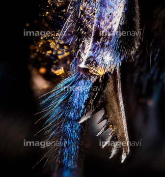
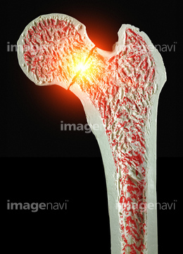
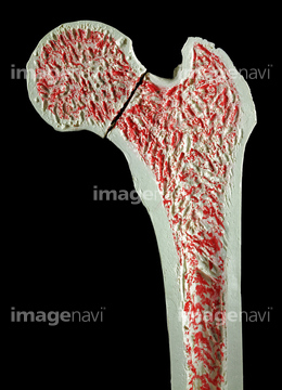
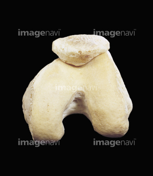
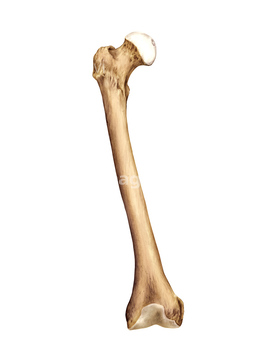
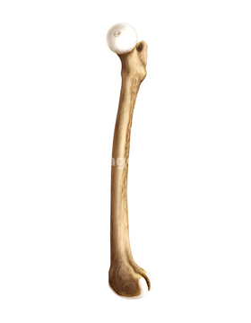
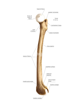
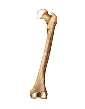
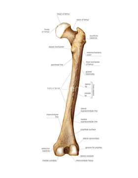
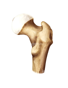
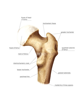
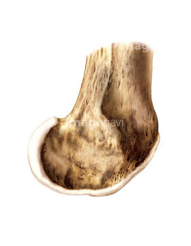
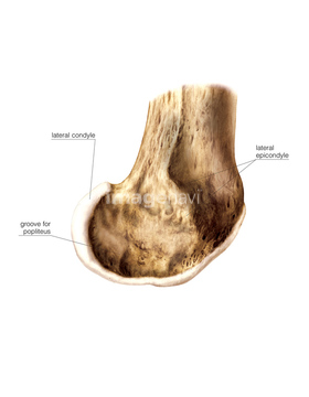
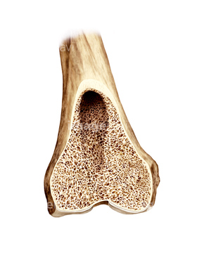
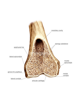
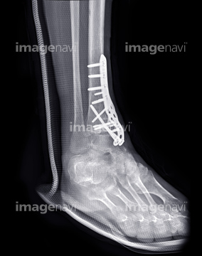
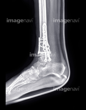
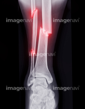
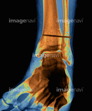
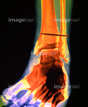
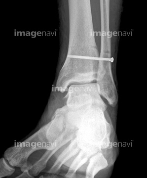
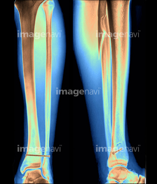
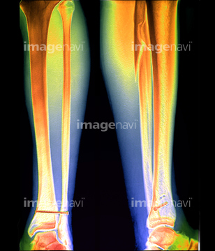
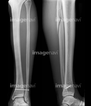
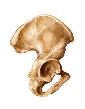
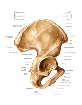
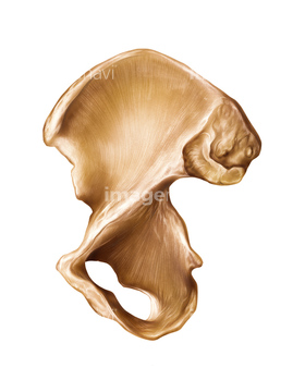
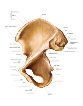
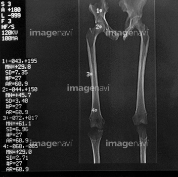
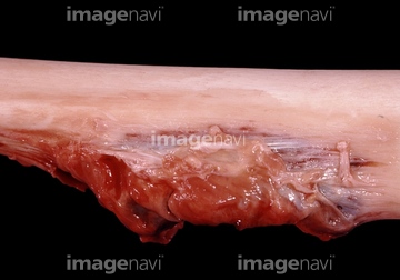
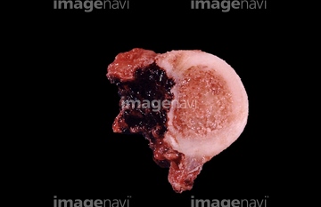
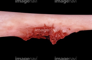
この検索結果には、Knee anatomy, artwork、Right femur proximal end from behind、Pinned broken leg, X-ray、Knee joint, light micrograph、Human leg and stiletto shoe, X-ray、Coloured MRI of a section through a knee jointなどが含まれています。
64225392

64225297
64225294
64225419
64225465
64225355
64225430
64225438
64224987
64070831
64070832
64070833
64070834
64070835
64070836
64224986
64225298
64224985
64070841
64070842
64071951
64071952
64071953
64100527
64070829
64070830
64225117
64040050
64070821
64070822
64074821
64225455
64224980
64070859
64070860
64225004
64106205
20527721
64225391
64225397
64225369
64056862
64225118
64072124
64071956
64225468

17206849
64146176
20529096
64059782
64060015
64225382
64147197
64147198
64147199
64225295
64225296
20559314
20559315
20559316
20559317
20559318
20559319
20559320
20559321
20559322
64010066
64012607
64012608
64012614
64012615
64071469
64071470

64083726
64060021
64059842
64059843
64059877
64225426
17201772
64083730
64225390
20527741
64149490
64149491
64149492
64149493
64149494
64149496
64225364
64225431
64225373
64225374
64225402
64013807
20559313
64243535
64010085
64071693
64071695
64071696
64187411
20562171
64093968
64093987
64101867
20527700
64071954
64071949
20569560
64149495
17237153
64184381
20566753
20559266
20559267
20559271
64109321
64225006
17218209
64188165
64225271
64078935
17206813
17211489
17200500
17261846
17261847
20562170
20527742
20569554
20573699
20566751
20564383
64245405
64245406
64225393

64262424
20559268
20559269
20559270
64225413
20569528
64104564
17237144
17237158
64225007
64225275
64225277
64050519
64070807
64070808
64070809
64070810
64070811
64070812
64070813
64070814
64070815
64070816
64225418
20566749
20566750
20566752
64262425
64262426
64262427
64262428
64262429
64262431
64070803
64070804
64070805
64070806
64219700
64219701
64219702
| 次ページ |
