HOME > 写真 > イラスト・CG > ビジネス > その他の職業
10,000件の写真素材が検索されました。
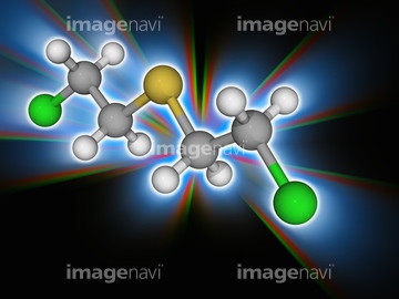
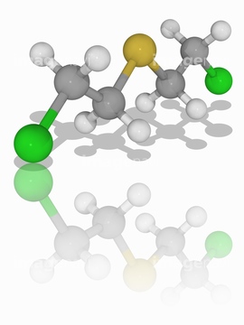
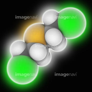
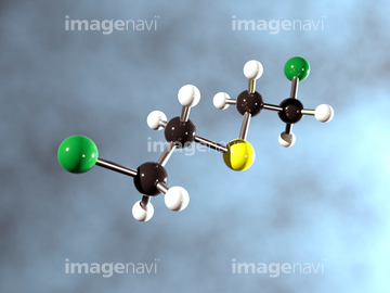
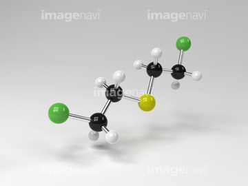
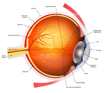
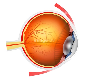
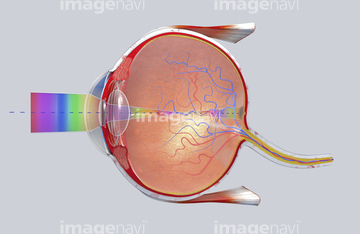
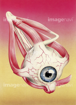
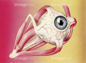
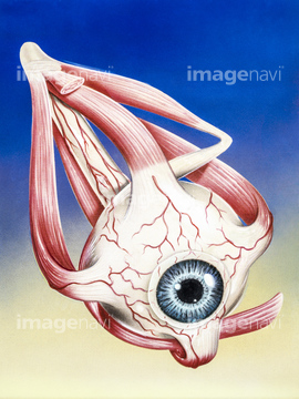
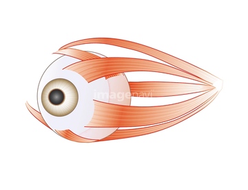
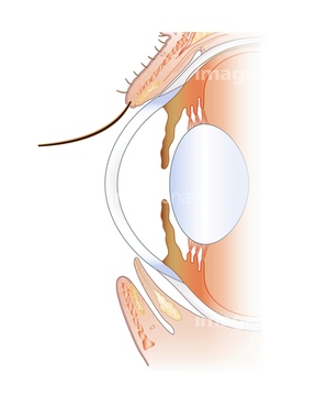
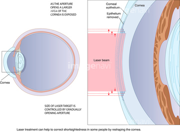
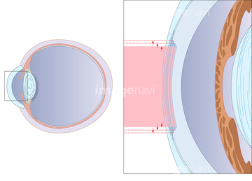
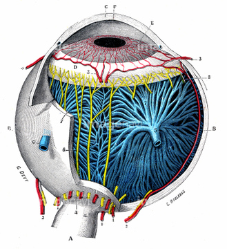
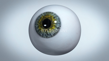
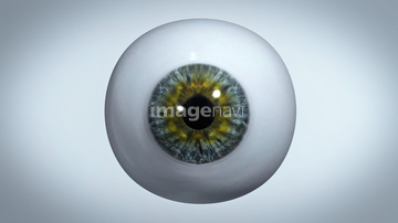
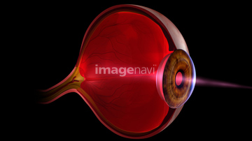
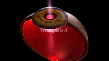
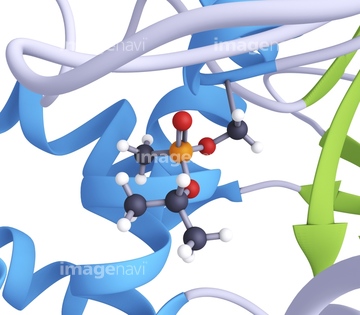
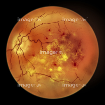
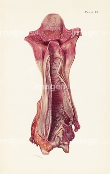
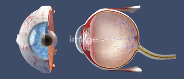
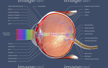
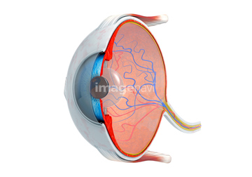
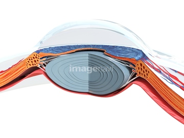
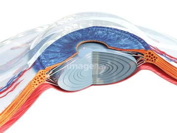
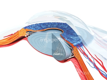
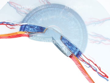
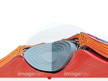
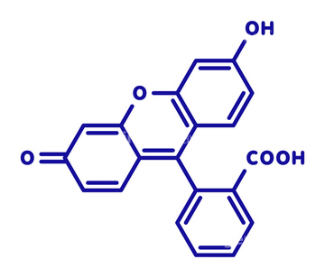
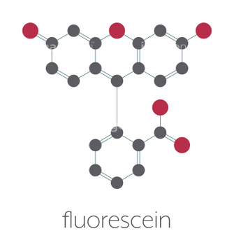
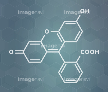
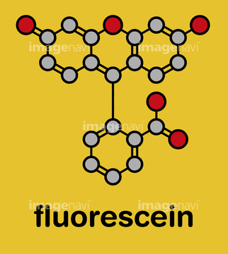
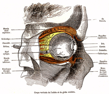
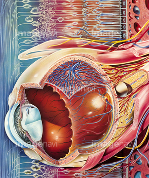
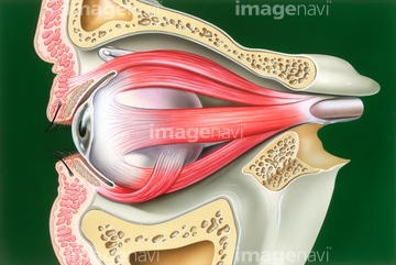
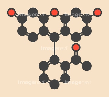
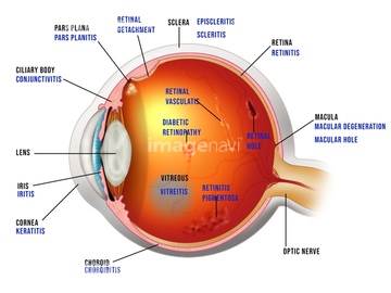
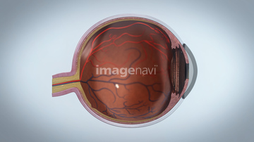
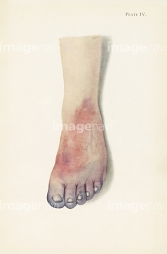
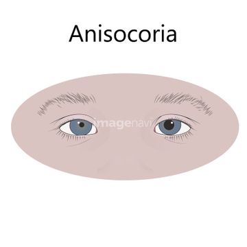
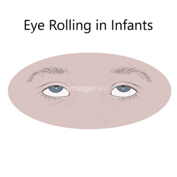
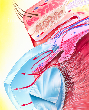
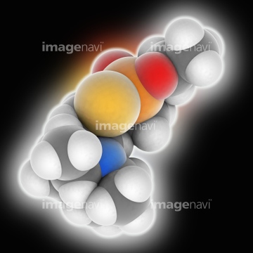
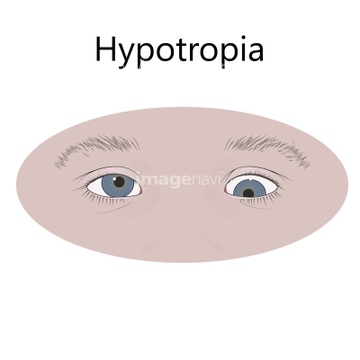
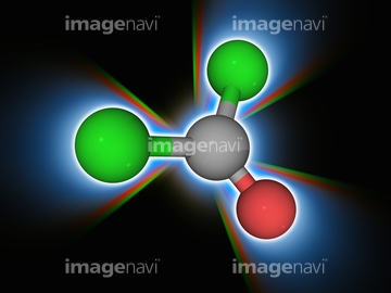
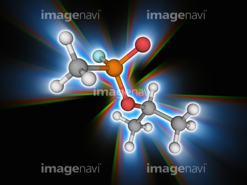
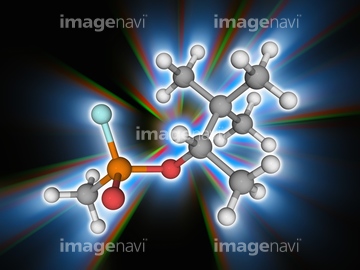
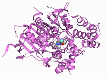
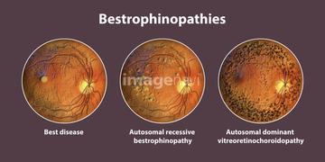
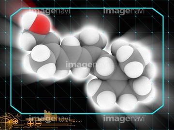
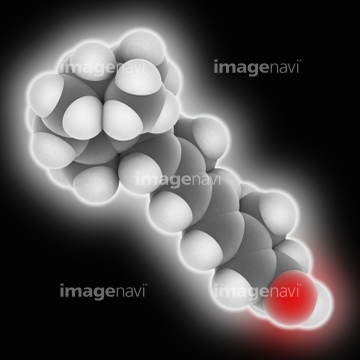
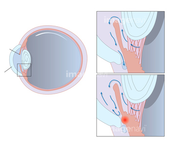
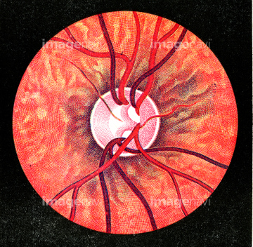
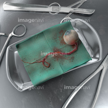
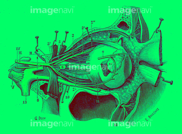
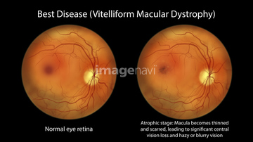
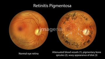
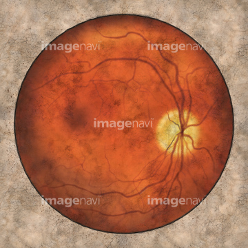
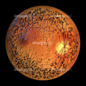
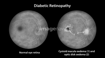
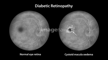
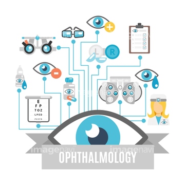
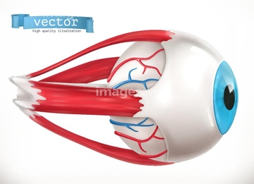
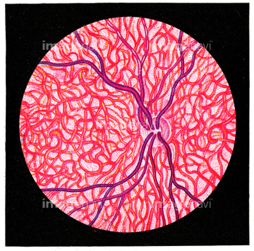
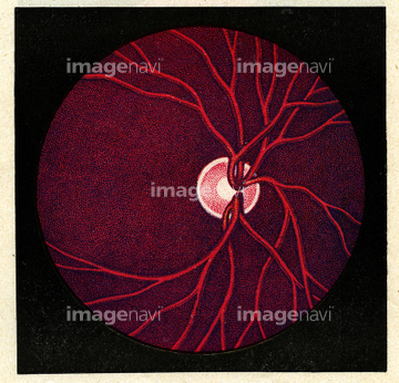
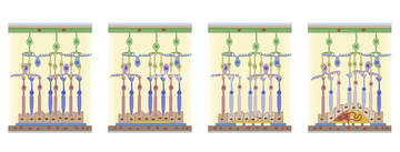
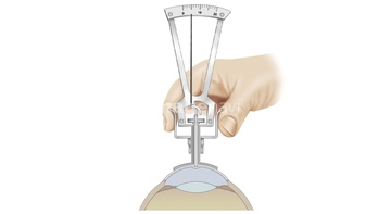
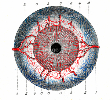
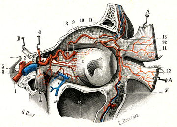
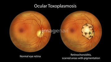
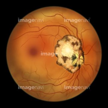
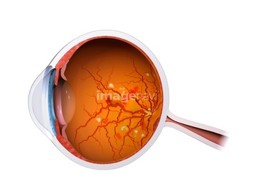
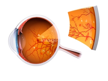
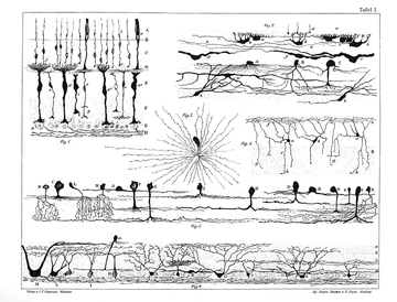
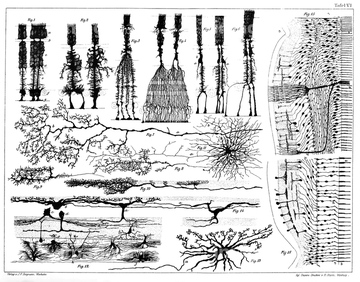
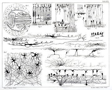
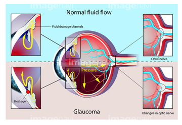
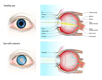
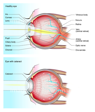
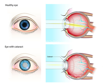
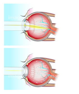
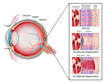
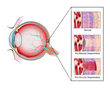
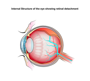
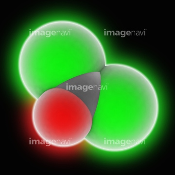
この検索結果には、Eye anatomy, illustration、Fluorescein fluorescent molecule、Eye anatomy, 19th century、Muscles of the eye, illustration、Eye diseases in slit lamp examination, illustratio…、Eye, illustrationなどが含まれています。

64102781
17299915
17284203
64252448
64264099
64252449

64089273

17227884
17227885
17284257
17284260
17294342
64207425
64207426
64207427
17227883
64125929
64045979
64045980
64256878
64249682
64249683
64245039
17294341
17294343
20528416
20537774
20537775
20537776
20537777
20537778
64175258
64175259
64175260
64175261
64178414
64207430
64119378
17246610
64249684
17299906
17299913
64089558
64103732
17299904
17299907
17299908
17299909
17299910
17299911
17299912
17259437
17259501
17259518
17258814
17258878
17258895
64169085
64169086
20574734
20528232
20528233
20528234
20528235
20528236
20528237
64142733
64103718
64044910
64257516
64252450
15802747
40523418
17299916

20574759
20574761
20574762
20574764
20574765
20574767
20574769
20574776
20574786
20560469
20560470
20560471
20560472
20560473
20560474
20560476
20560477
20560478
29083796
29457074
64257514
64179379
64256877
64257515
64209383
20574735
64258094
64258095
64258096
17204383
64103026
29110916
29110917
| 次ページ |
