HOME > 写真 > イラスト・CG > 人物 > 男性
10,000件の写真素材が検索されました。
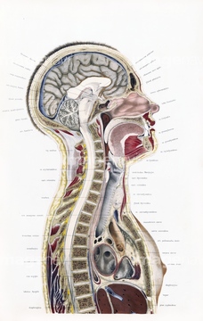
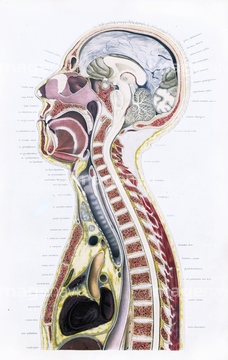
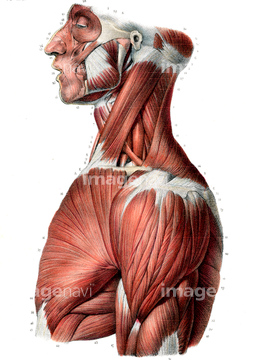
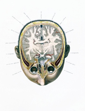
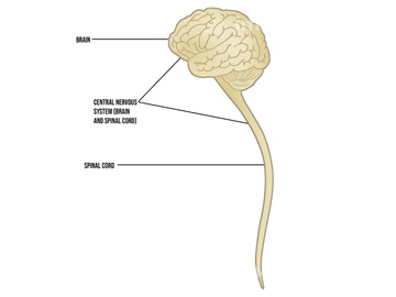
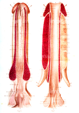
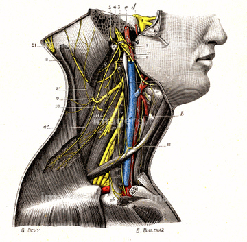
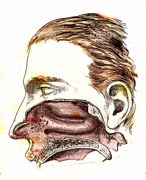
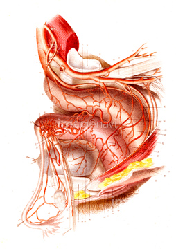
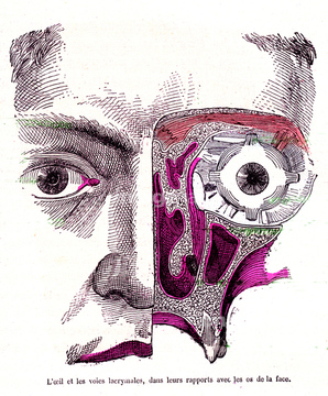
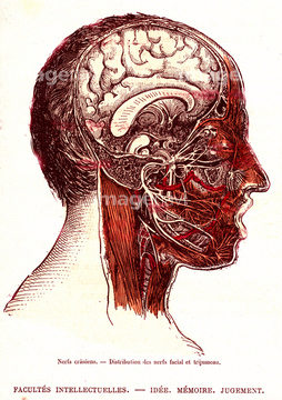
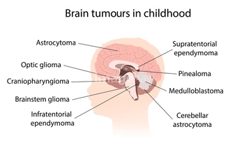
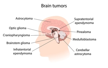
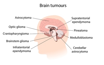
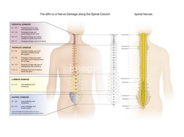
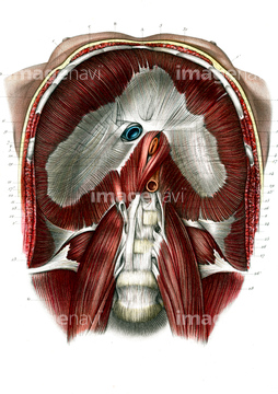
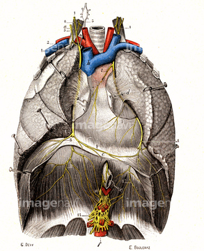
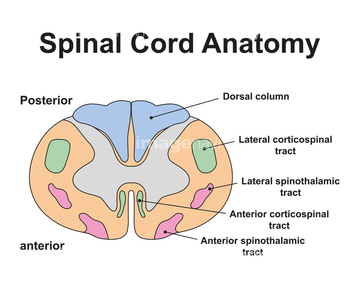
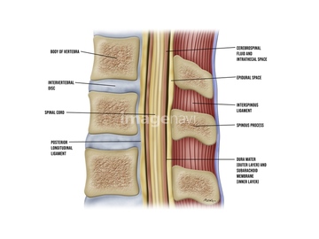
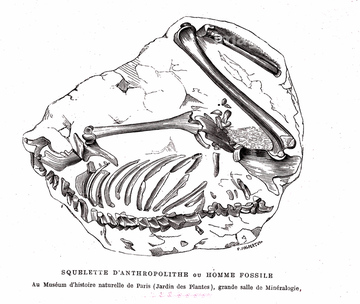
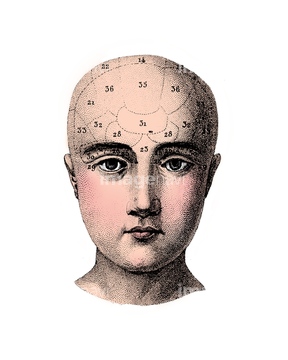
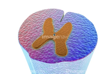
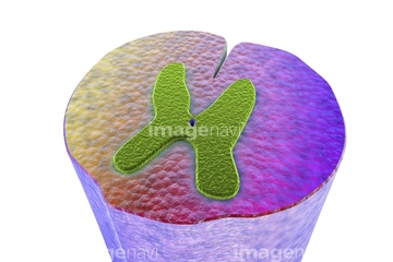
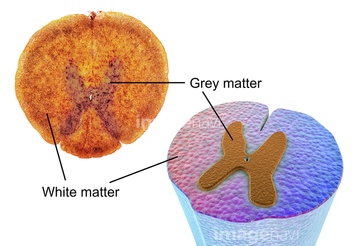
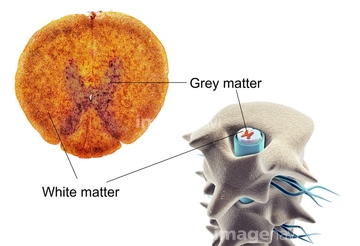
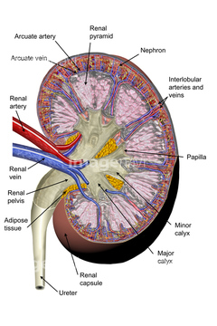
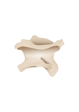
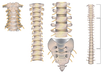
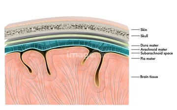
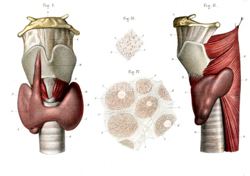
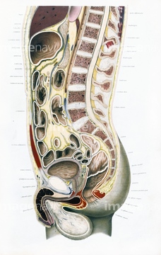
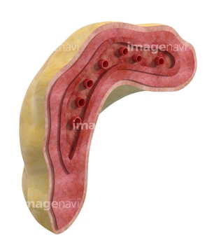
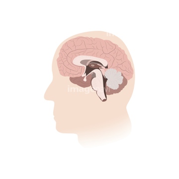
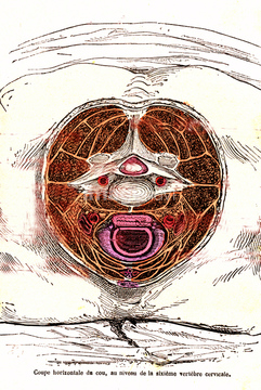
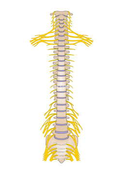
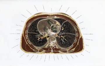
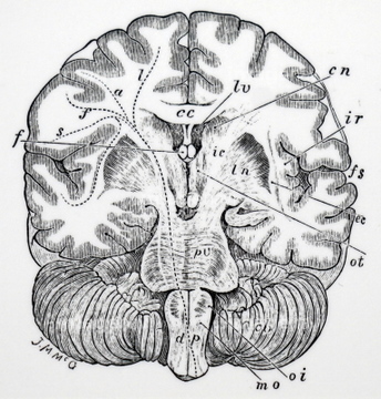
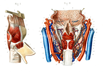
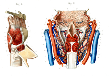
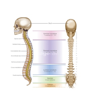
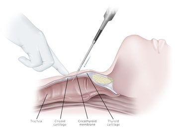
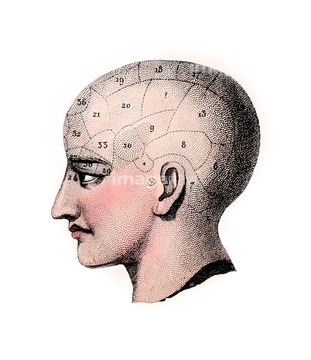
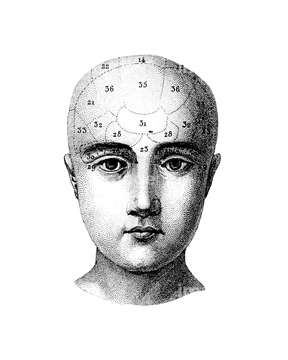
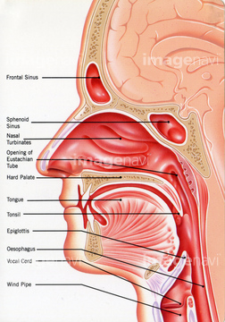
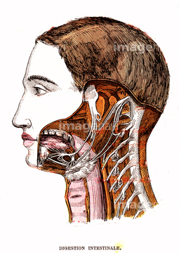
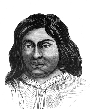
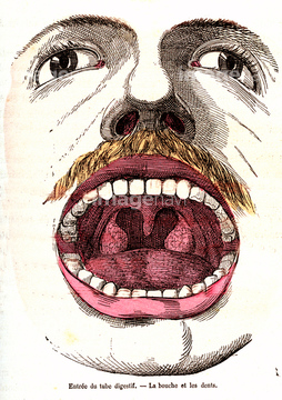
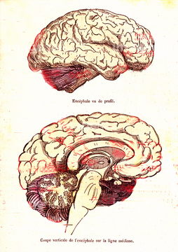
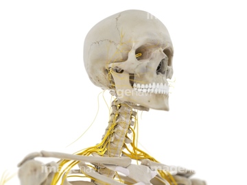
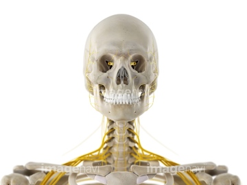
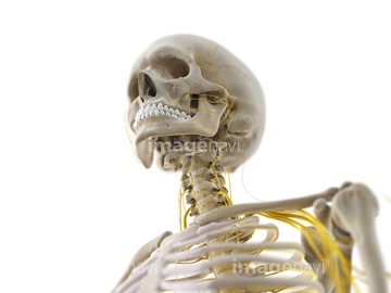
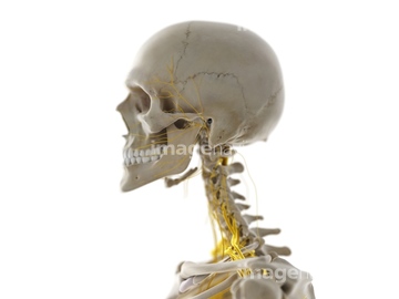
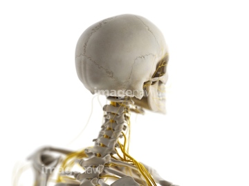
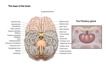
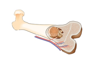
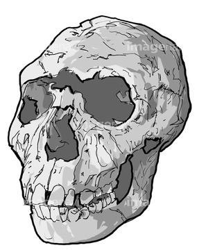
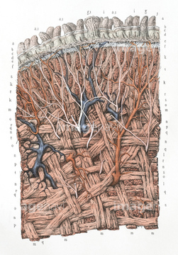
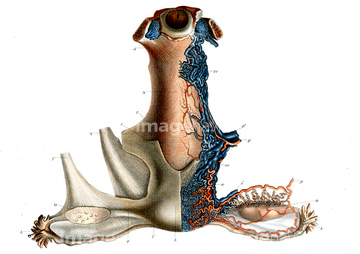
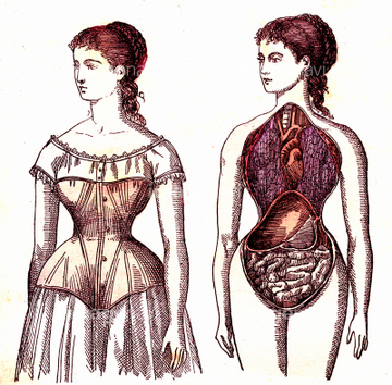
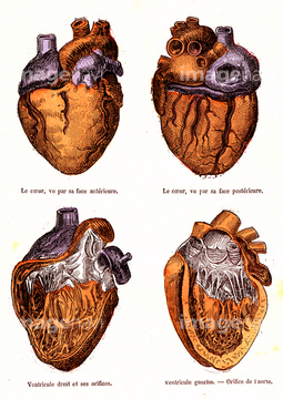
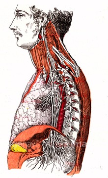
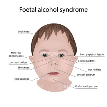
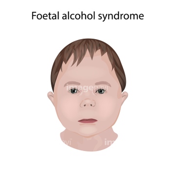
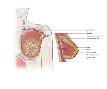
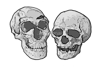
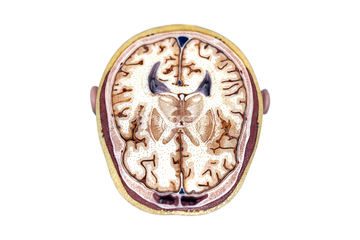
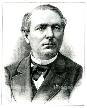
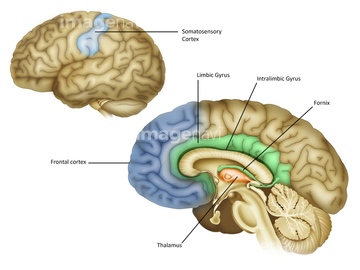
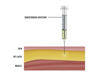
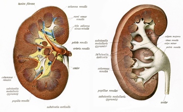
この検索結果には、Phrenic nerves, illustration、Spinal cord anatomy, illustration、Human fossil, 19th century illustration、Phrenology head, illustration、Spinal cord cross-section, illustration、Kidney anatomy, illustrationなどが含まれています。
64110023
64177872

64110026
64257573
64109999
64110001
64110029
64177922
64177892
20547112

64126082
64109997
64109998
64110000
64110005
64110010
64110021
64110022
64110033
64110035
64110037
64110052
64110053
64110038
64110041
64257571
64257572
20571228
64177873
64124771
64131930
64241930
64232261
64125581
64110002
64144311
20547079


20547069

20547072

20547073
20547074
20547075

20547076
20547077
20547078
20547092
20547095
20547096

20547097
20547098
20547099

20547100

20547101

64244370
64261066
64110011
64110012

64110039

64110040
64110049
20547549
64126081
64124793
64124798
64207401
64217786
64088457
64068189

64177897
20519472
20519473
20519474
20519475
20519476
64232089
64085602
64191005
64063127
64064231
64064232
64064257
64064258
64064259
64232075
64168659
64088464
64110013

64110015
64110019
64110042
64110047
64241938
64241957
64251889
64217384
64232077
64177909
20537918
20547164
20547547
64177876
64177894
64183409
| 次ページ |
