HOME > 写真 > 科学・テクノロジー > 科学 > DNA・細胞
10,000件の写真素材が検索されました。
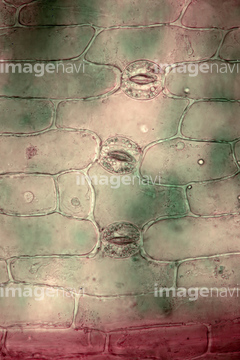
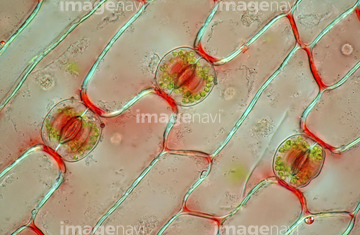
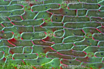
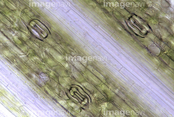
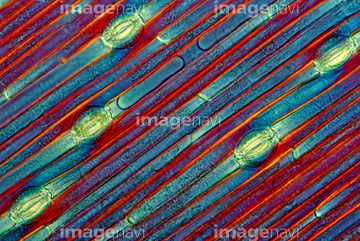
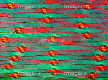
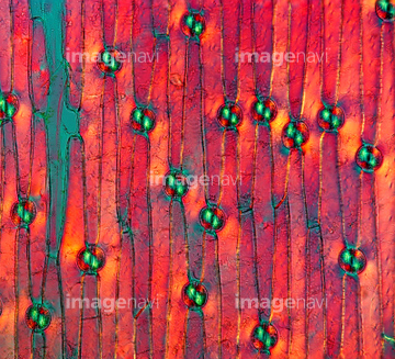
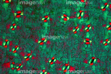
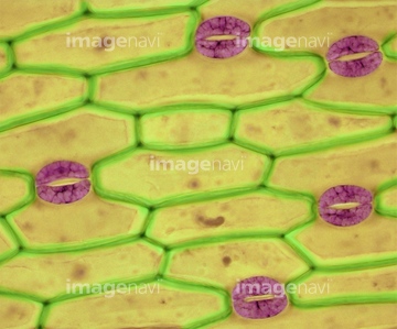
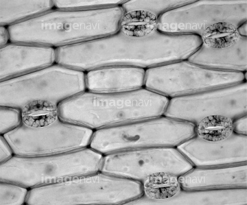
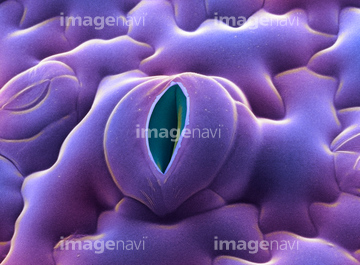
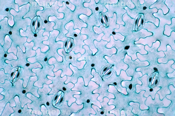
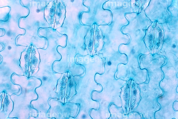
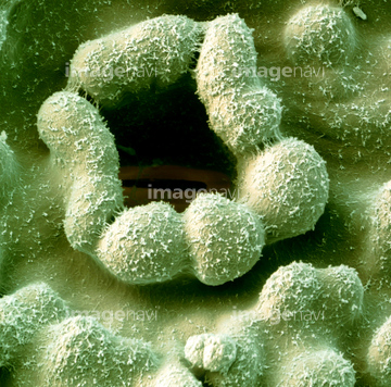
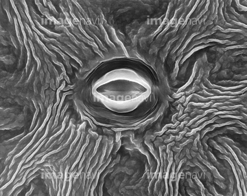
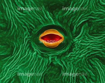
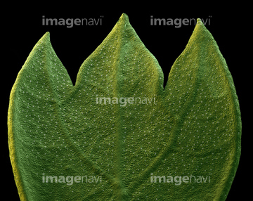
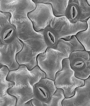
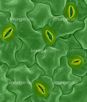
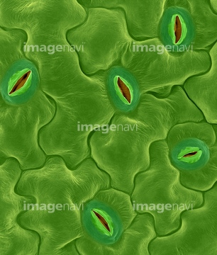
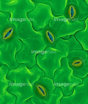
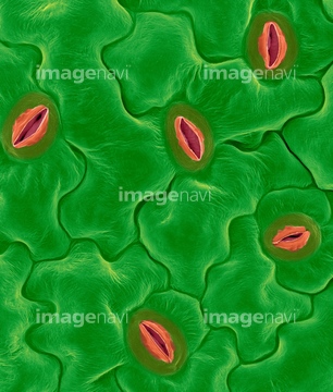
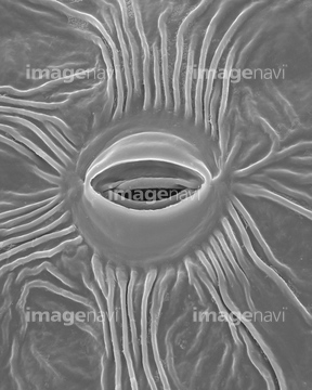
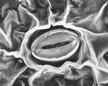
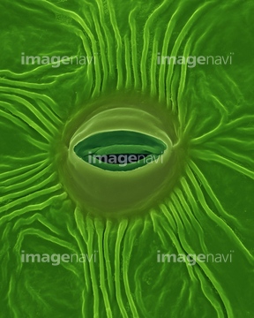
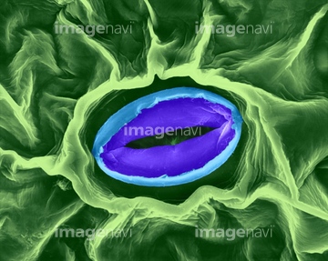
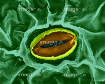
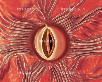
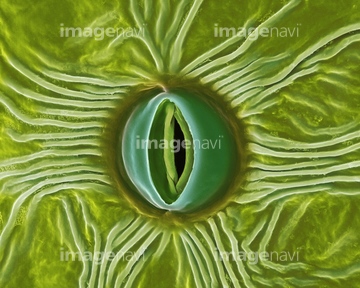
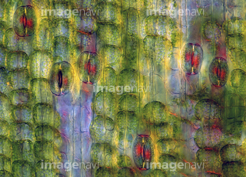
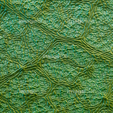
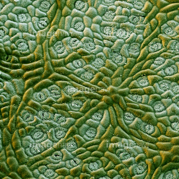
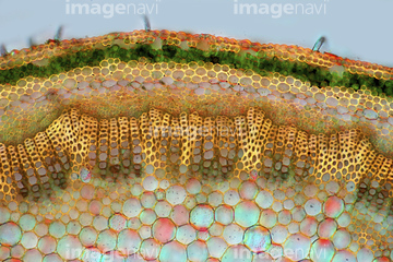
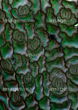
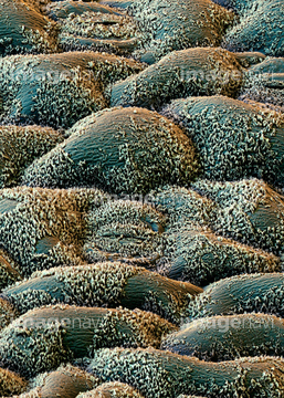
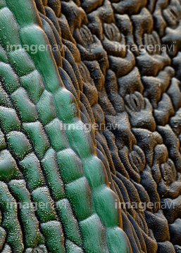
この検索結果には、Fern leaf stomata, light micrograph、Tulip leaf stomata, light micrograph、Closed stoma, SEM、Open stoma, SEM、Leaf epidermis, light micrograph、Tulip stomata, light micrographなどが含まれています。
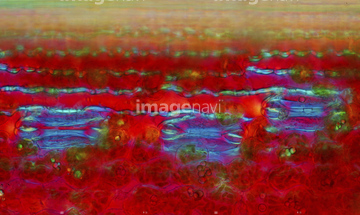
64259886
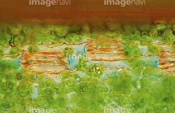
64259882
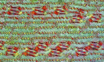
64259884
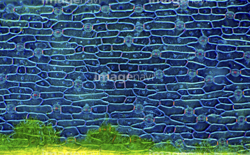
64259885
64259887
64259888
64259889
64259890
64259891
64259974
64259966
64259968
64259969
64259973
20542488
64259972
64259967
64089408
64089438

64153105
64089521
64098748
64098749
64099743
64105240
64105241
64105251
64105268
64105281
64224866

64145495
64145512
64155055
64208905
64208906
64208907
64255079
64255080
64005703
64005706
20530800
20542520
20542521
64098756
64105252
64105253
64089520
64255901
64099687
64105289
64105290
64105291
64005705
64090375
64090376
64005716
64005717
64005722
64006295
64006296
64162969
64087657
64250245
64089505
64089506
64089507
64005704
64005718
64114726

64089399
64213957
64213976
64157866
64140430
64195202
64195203

64195204
64195205
64195206
64195208
64089522
64089523
64261348
64261349
64261351
64261354
64056494
64005633
64150985
64137210
64199842
64151007
64137288
64137289
64137290
64137291
64117900
64259862
64259881
64259905
64259908
64005681
64005682
64005683
64005684
64138238
64138259
64197477
64261350
64089396
64089397
64089398
64090470
64123982
64129207
64005637
64259906
64259907
64259922
64195207
64119713
64220277
20547148
| 次ページ |
