HOME > 写真 > 科学・テクノロジー > 科学 > DNA・細胞
10,000件の写真素材が検索されました。
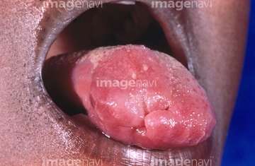
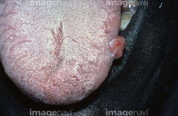
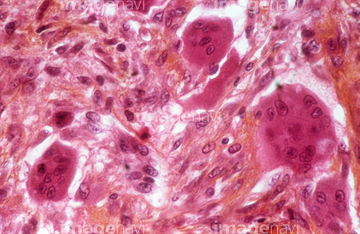
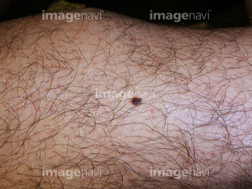
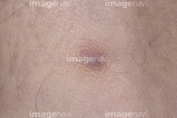
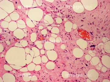
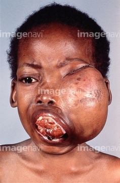
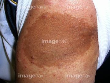
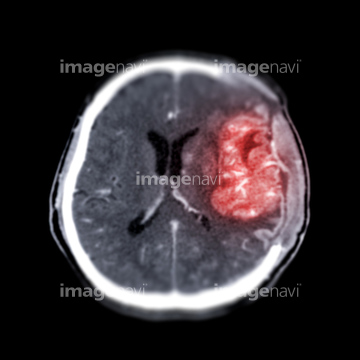
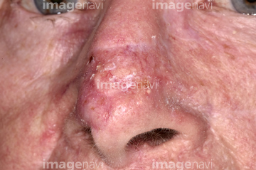
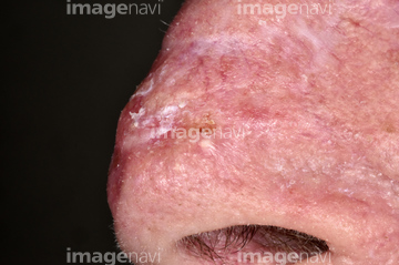
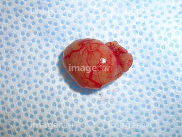
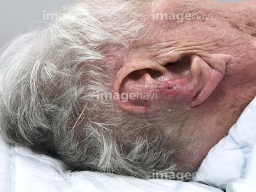
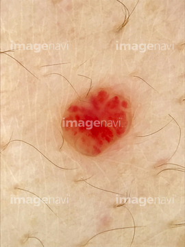
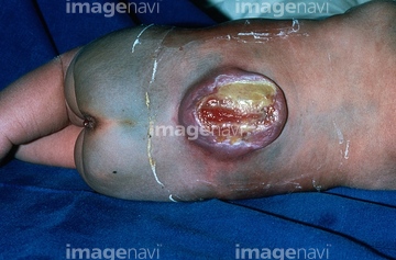
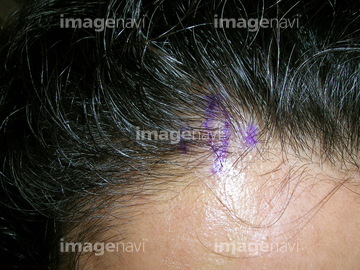
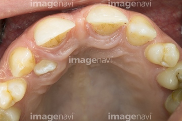
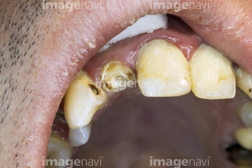
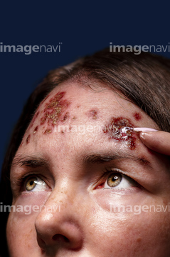
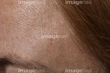
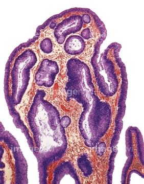
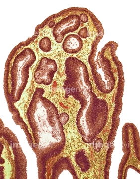
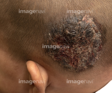
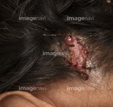
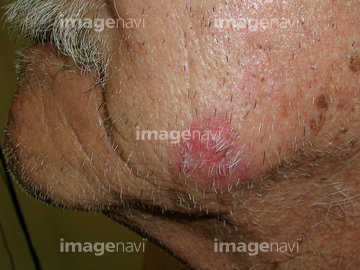
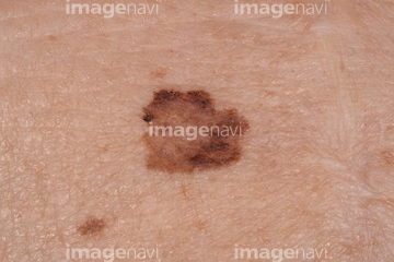
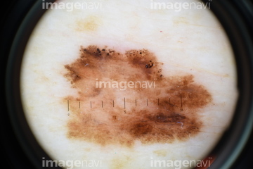
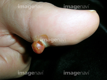
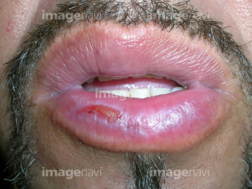
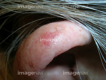
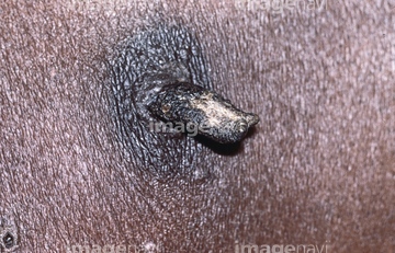
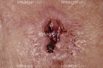
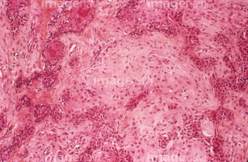
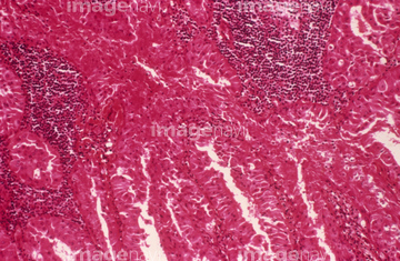
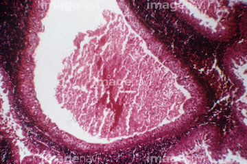
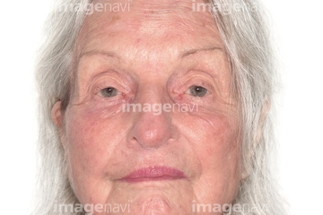
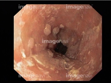
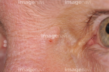
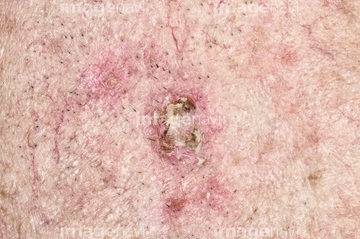
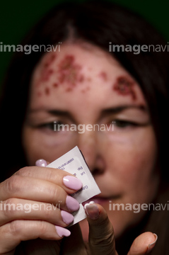
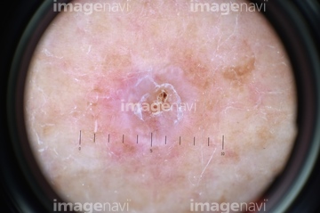
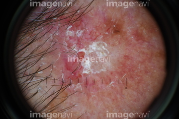
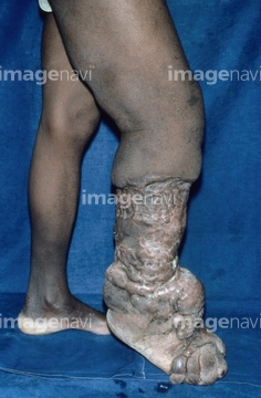
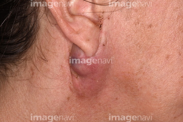
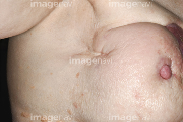
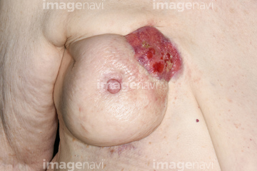
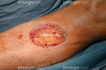
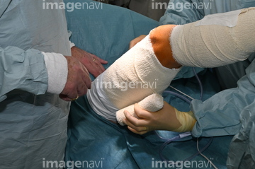
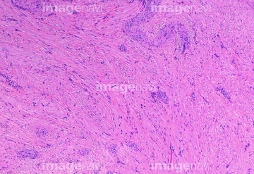
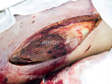
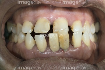
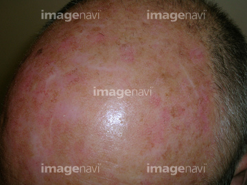
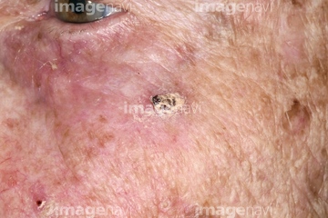
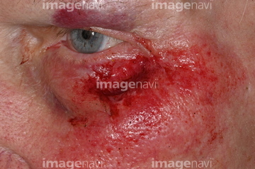
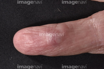
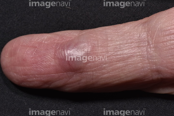
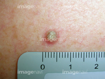
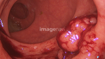
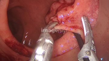
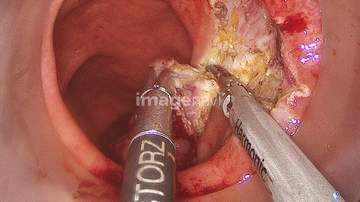
この検索結果には、Secondary oral cancer、Fibrosarcoma, light micrograph、Open wound after small bowel surgery、Wound after surgery to remove basal cell carcinoma、Campbell de Morgan spot, dermoscopy、Lumbar meningiomaなどが含まれています。
64264747
64264650
64264697

64253948
64253949
64252672
64252673
64264696
64264818
64092796
64252731
64204903
64264741
64252689
64254453

64159301
64159302
64177832
64177833
64184301
64204928
64204929
64210363
64210364
64252732
64253410
64253411
64253412
64253414
64244446
64244447
64213166
64265715
64263273
64252651
64264618
64204878
64204913
64244463
64244461
64254472
64254473
64254476
64264999
64152361
64155645
64213101
64252696
64264742
64253979
64126119
64204935
64265699
64092845
64092846
64155670
64155680
64155695
64155696
64264904
64177831
64204905
64210365
64262952
64262992
64262994
64264689
64205890
64225543
64225533
64225826
64225827
64253396
64155657
64155701
64204313
64222507
64252685
64254439
64254463
64254477
64264682
64264806
64252690
64265294
64205894
64205902
64096091
64204288
64207288
64207294
64213171
64225824
64225825
64225842
64225843
64257279
64244388
64252662
64252683
64253256
64253257
64253986
64254458
64254474
64264686
64204931
64204932
64225752
64212404
64217943
64254461
64244472
64219329
64204930
64207292
64225835
64264688
64213104
64263907
64263908
64126656
64152948
64252684
64129320
64129324
64244414
64225665
64225666
64265691
64204696
64204697
64257266
64259839
64259840
64259842
64259843
64259844
64198943
64188409
64221755
64172411
64244413
64188267
64172367
64244407

64244408
64244409
64195721
64195722
64167391
64264704
64254459
64173396
64152362
64152585
64201878
64205330
64205331
64230414
64230415
64265698
| 次ページ |
