HOME > 写真 > 科学・テクノロジー > 科学 > DNA・細胞
10,000件の写真素材が検索されました。
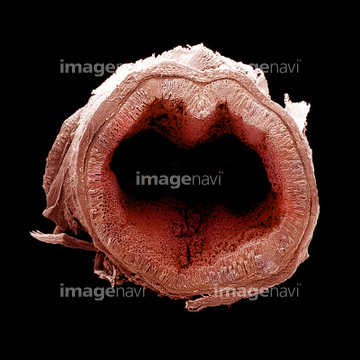
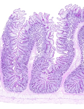
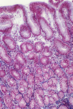
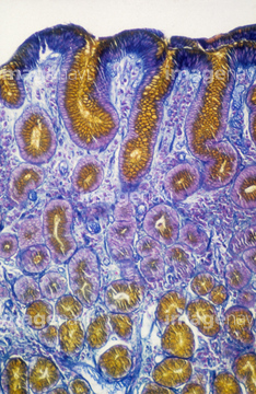
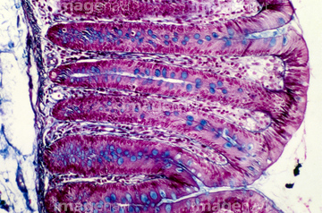
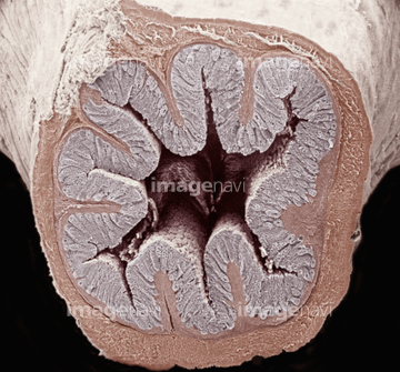
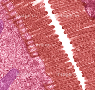
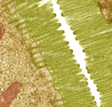
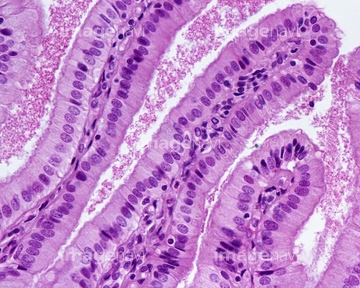
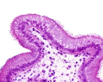
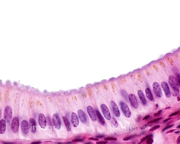
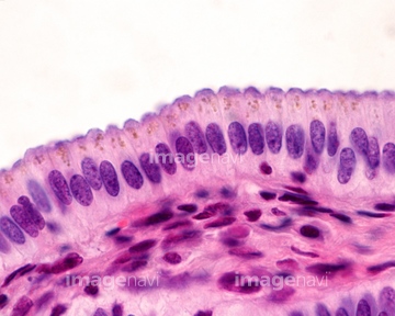
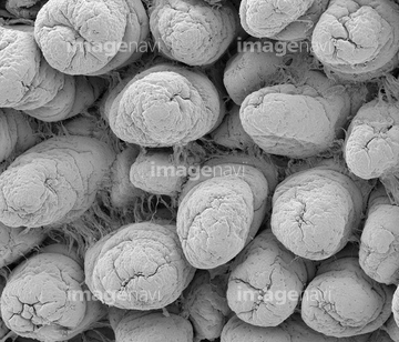
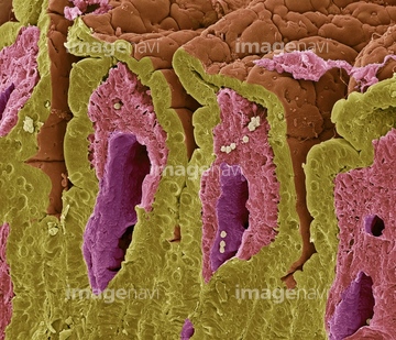
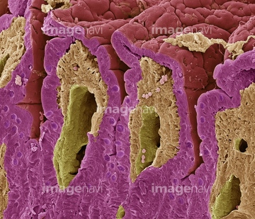
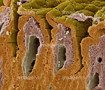
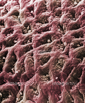
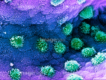
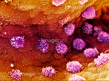
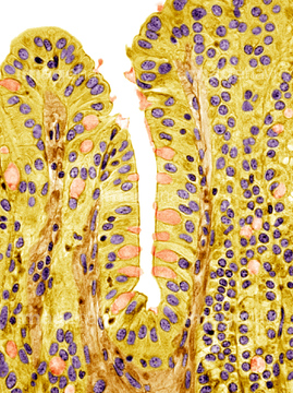
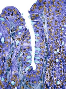
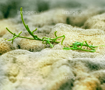
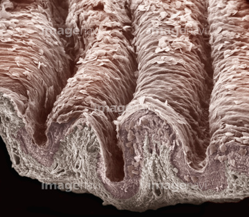
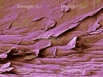
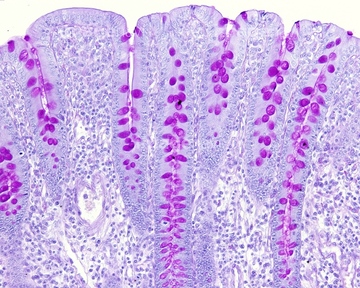
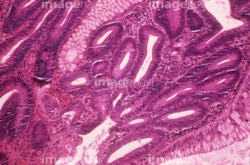
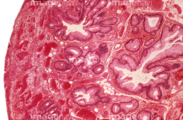
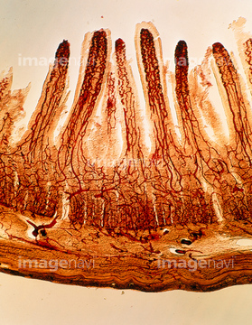
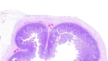
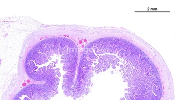
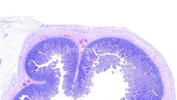
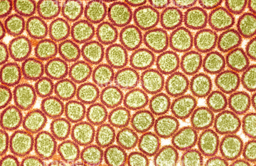
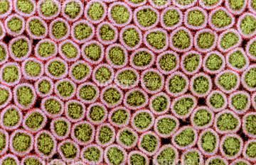
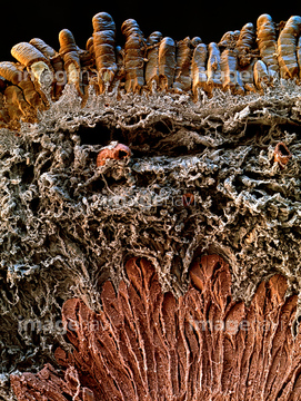
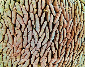
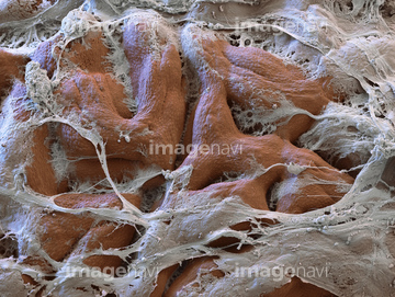
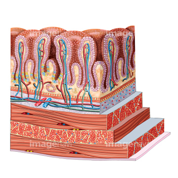
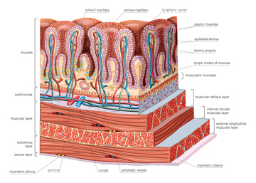
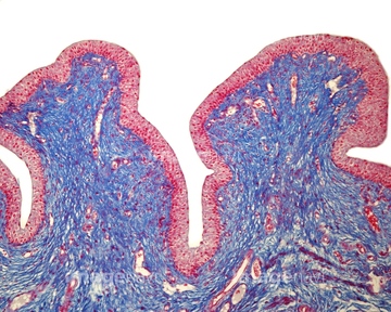
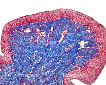
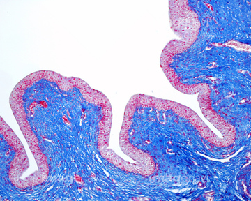
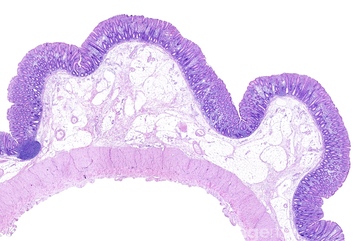
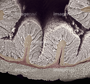
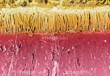
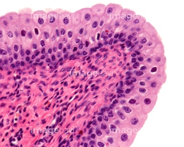
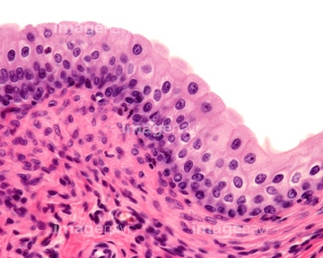
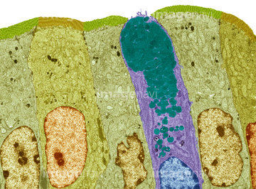
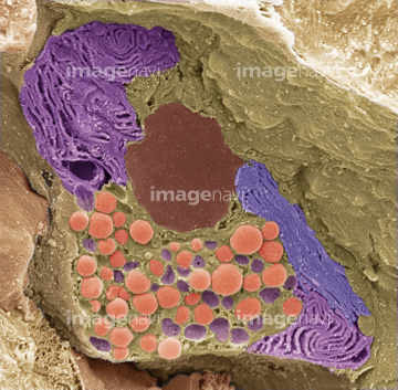
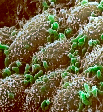
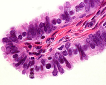
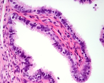
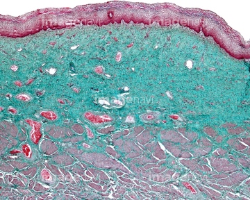
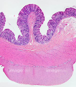
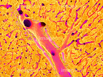
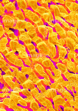
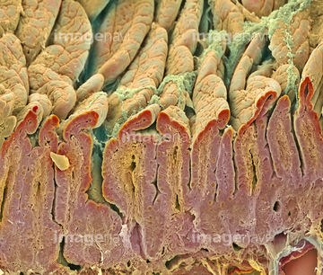
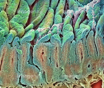
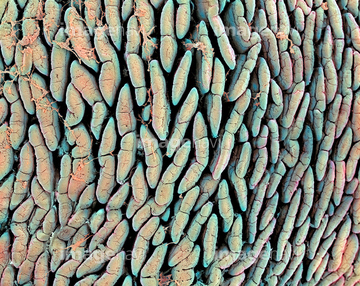
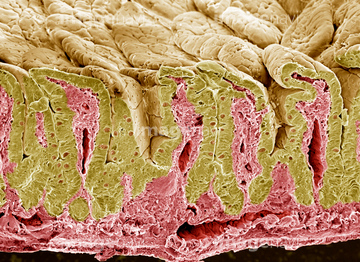
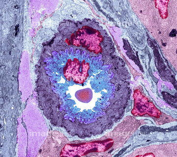
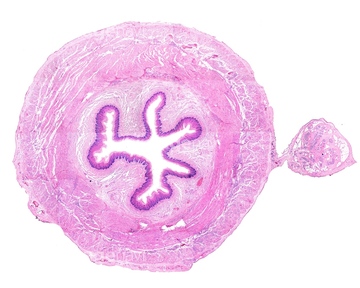
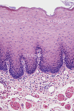
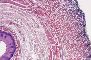
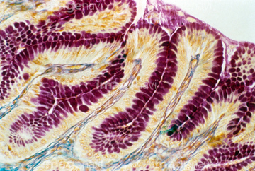
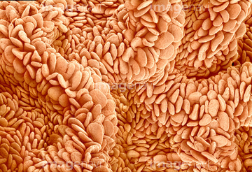
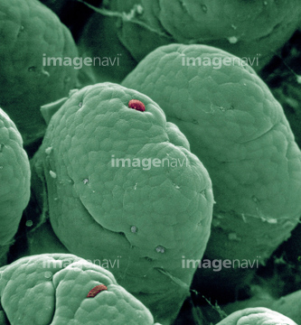
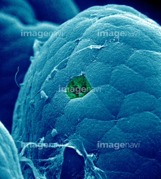
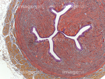
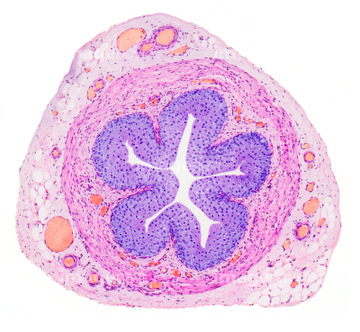
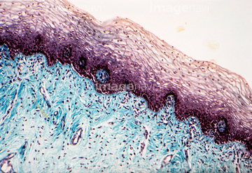
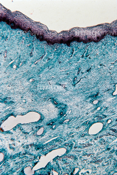
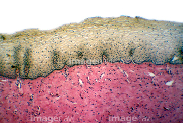
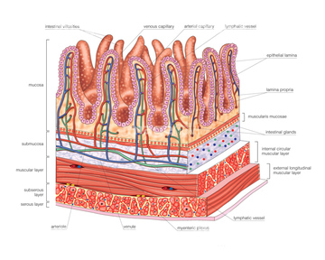
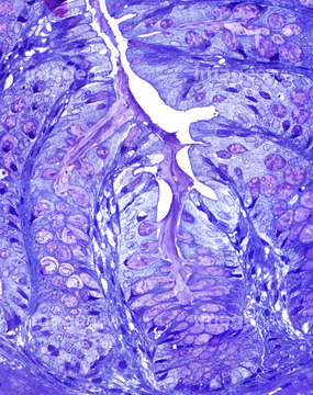
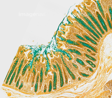
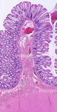
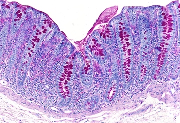
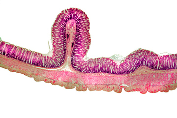
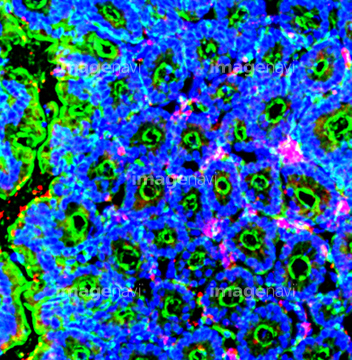
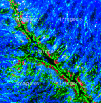
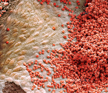
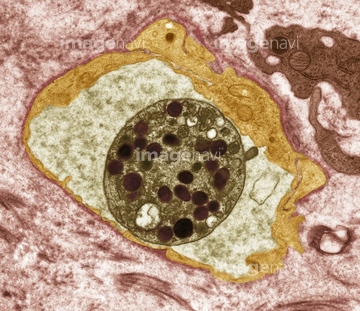
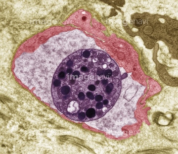
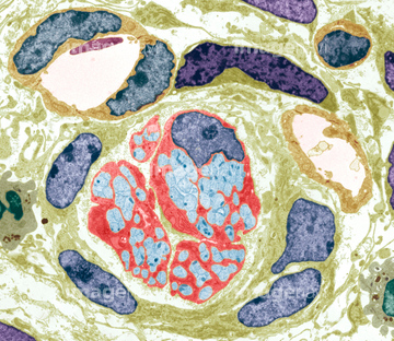
この検索結果には、Surface of the stomach、Colon goblet cells, SEM、Small intestine lining, light micrograph、Parasites in large intestine, SEM、Stratified squamous epithelium, vagina、Vaginal lining, SEMなどが含まれています。
64008751
64008746
64008744
64008745
64225131
64206819
64073272
64073273
64073274
64073275
64073276
64225189
64071989
17202471
17202472

64146135

64146275
64221011
64066084
64196734
64190237
64008158
64074891
64073988
64071892
64071893
64152547
64192859
64192866
64192886
64225092
64021658
64021659
64055754
64215656
64082486
64082487
64073315
20542530
20544099
64197486
64197489
64199159

64225140
64073304
20523344
20523360
64198564
64084027
64213913
64213914
64213915
64060825
64060826
64065178
17201424
64176856
64176857
64176858
20507330
20507334
64071850
64071851
64196735
64077368
64061536
64075661
64056271
64265102
64265137
64265142
64066076
64061534
64061535
64063559
64063560
64063561
64073344
64021586
64021585
64021605

64206746
64206747
64196715
64088343
64002984
64002985
64177447
64090172
64076362
64076363
64021539
64021540
64021541
64021571
64060203
64060204
64205896
64205907
64085267
64197478
64235009
64235010
64071988
64008180
64073608
64049910
64049911
64088791
64093995
64063011
64063015
64065986
64077369

64225083

64225133
64234280
64225192
64155017
64225129
64071876
64071877
64090158
64090187
64090463
64106063
64085258
64214176
64057542
64072074
64136447
64234508
64045958
64204230
64204232

64037575
64037576
64076022
64076023
64071833
64071834
64234509
64234510
64234842
64234476
64234477
64234478
64234479
17200013
| 次ページ |
