HOME > 写真 > 科学・テクノロジー > 科学 > DNA・細胞
10,000件の写真素材が検索されました。
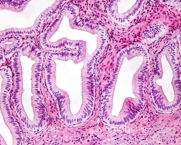
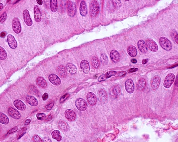
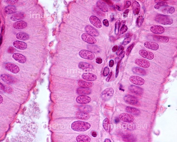
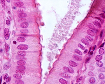
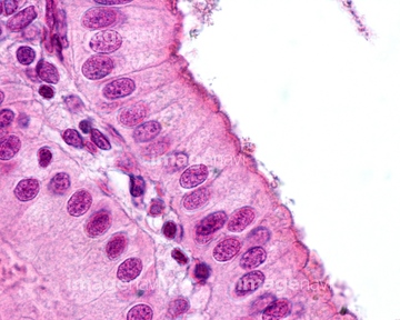
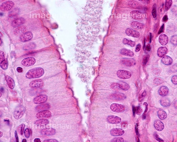
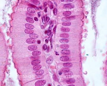
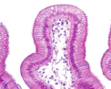
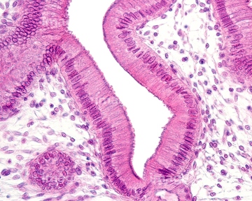
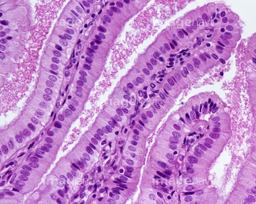
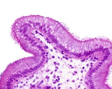
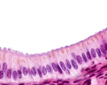
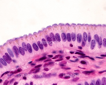
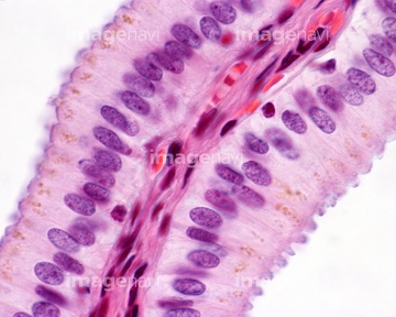
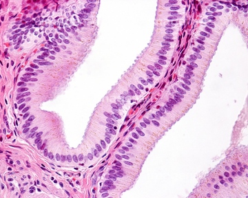
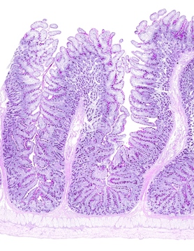
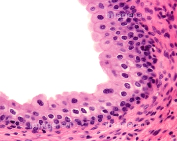
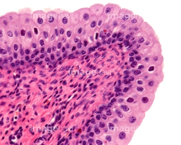
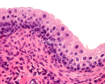
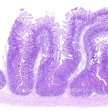
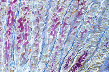
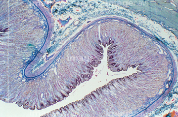
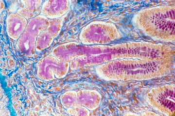
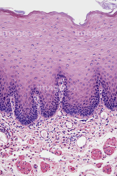
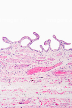
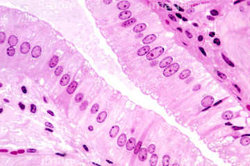
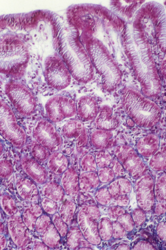
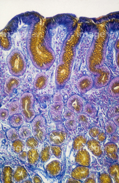
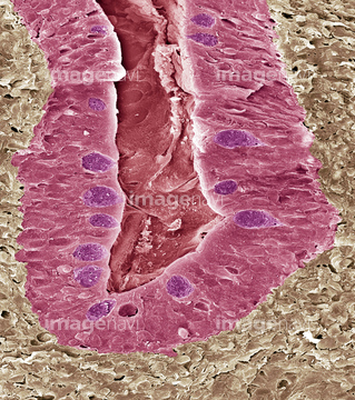
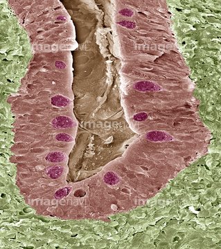
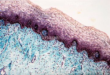
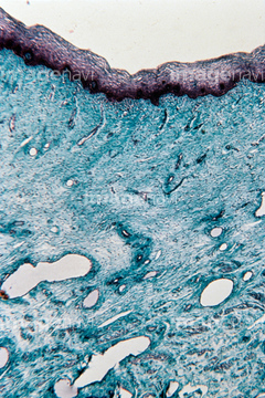
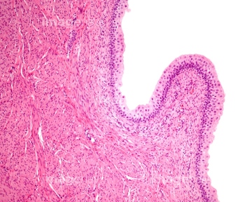
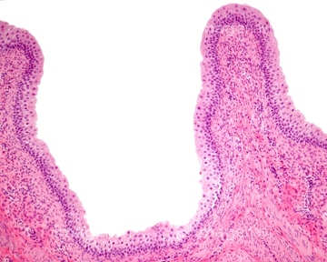
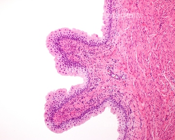
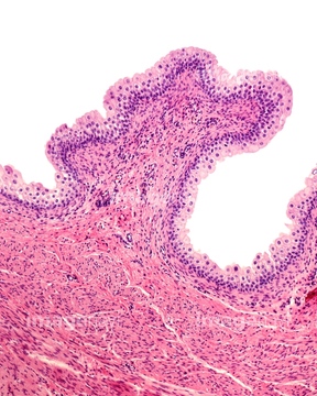
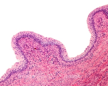
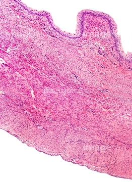
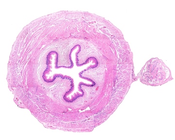
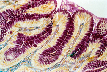
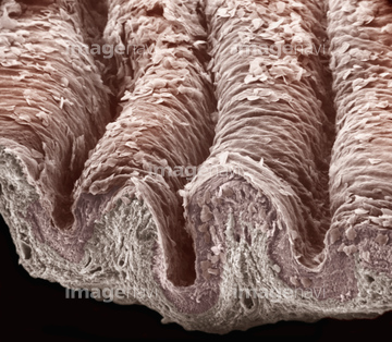
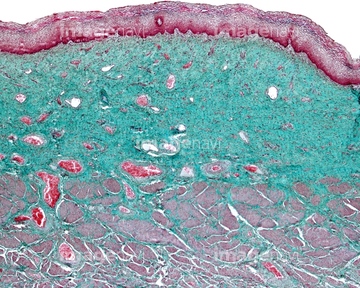
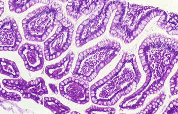
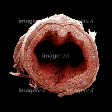
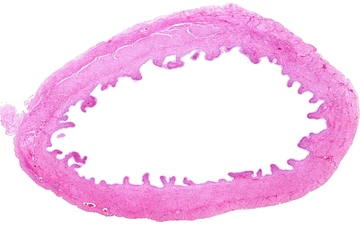
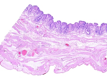
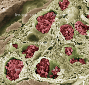
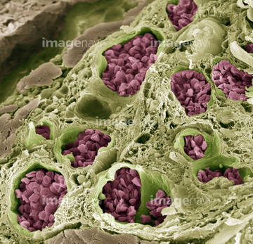
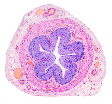
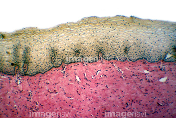
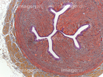
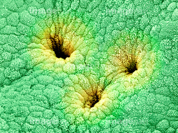
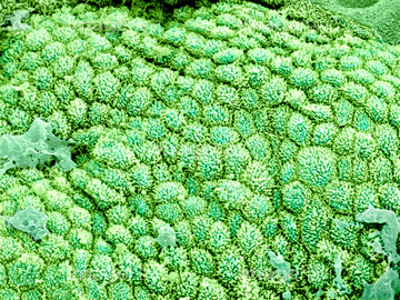
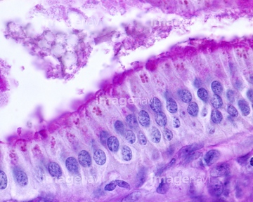
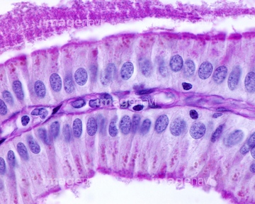
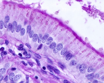
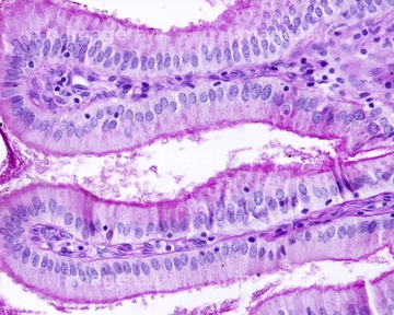
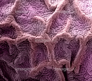
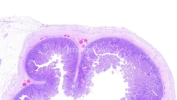
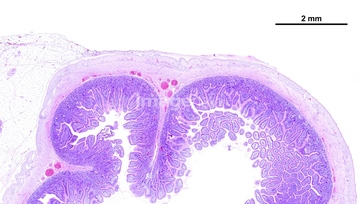
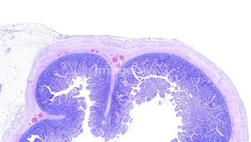
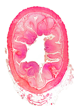
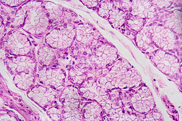
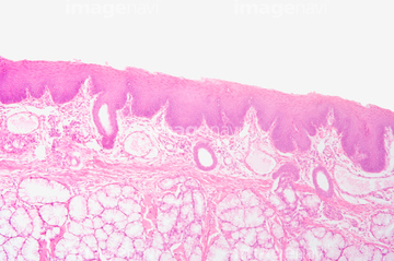
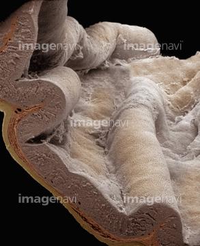
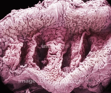
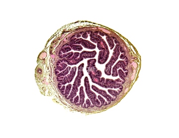
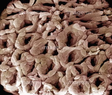
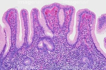
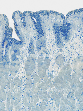
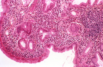
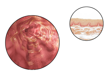
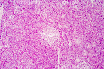
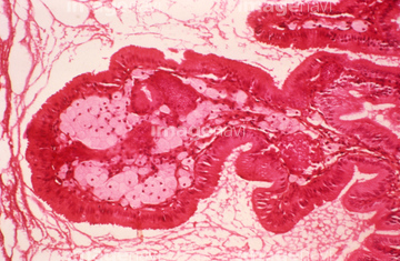
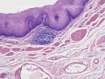
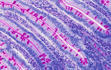
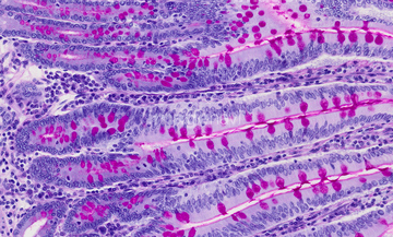
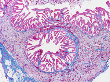
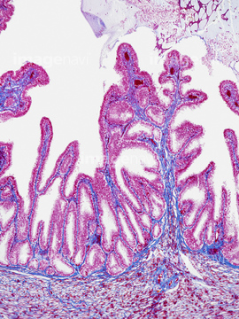
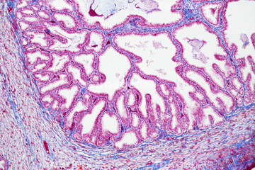
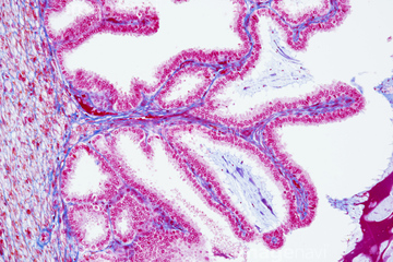
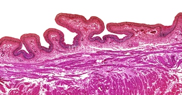
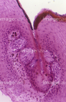
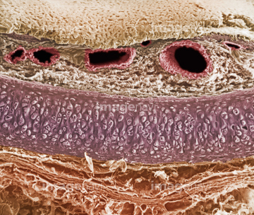
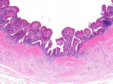
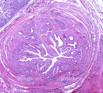
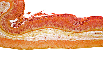
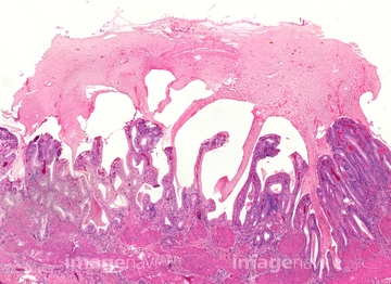
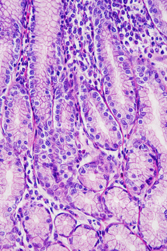
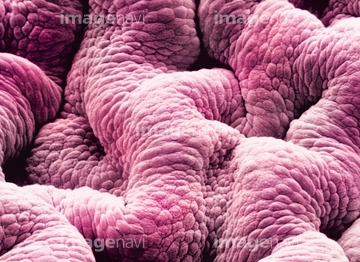
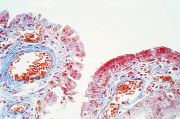
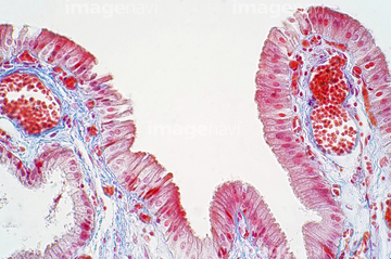
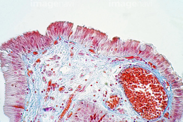
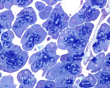
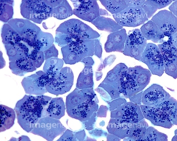
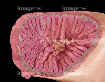
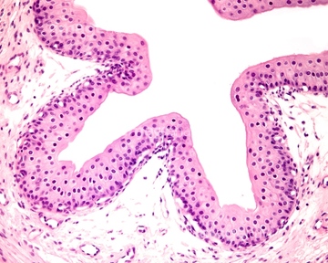
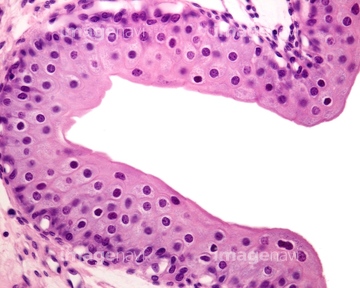
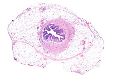
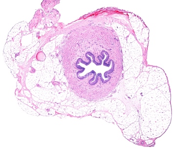
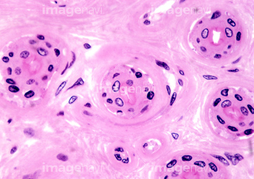
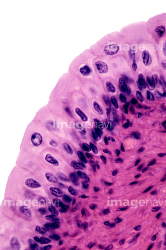
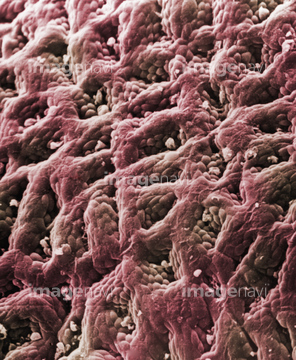
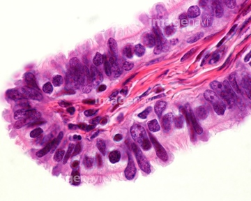
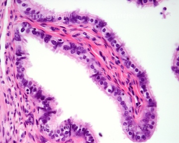
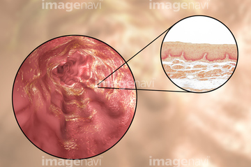
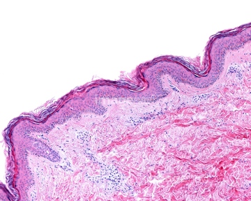
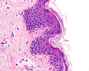
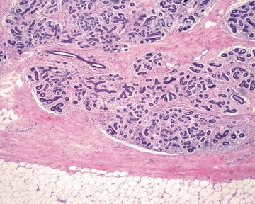
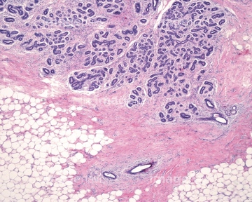
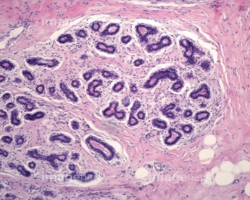
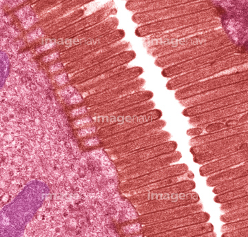
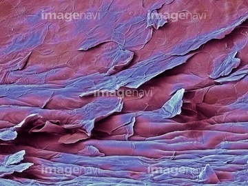
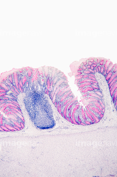
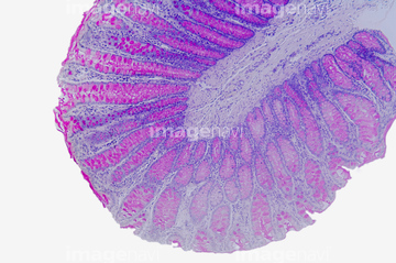
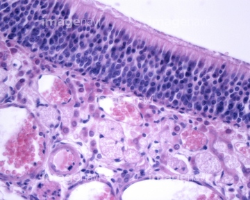
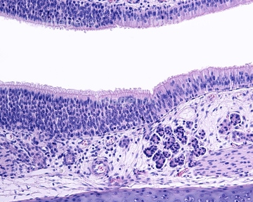
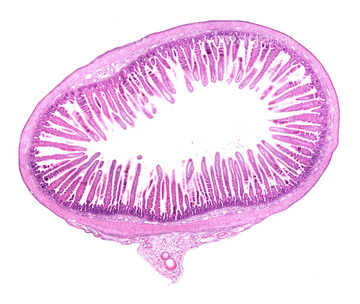
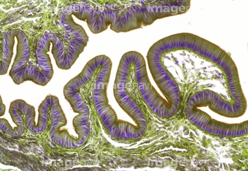
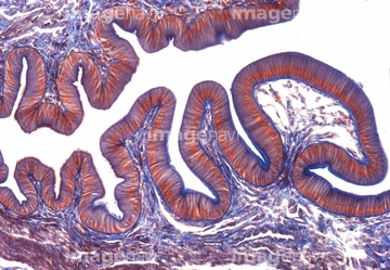
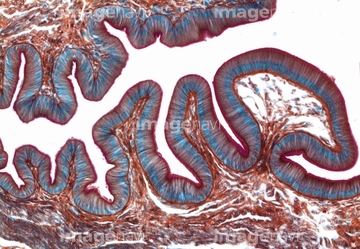
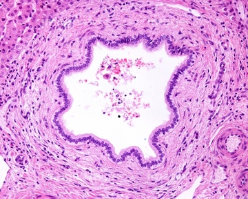
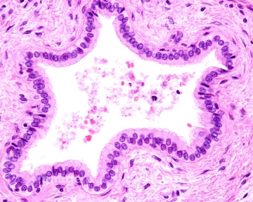
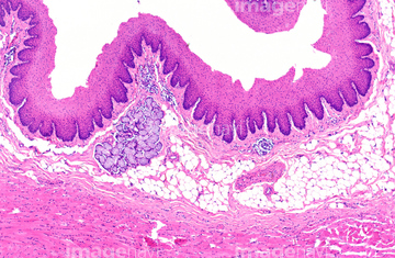
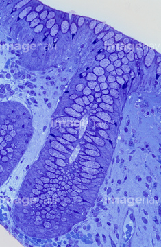
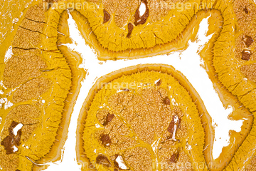
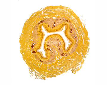
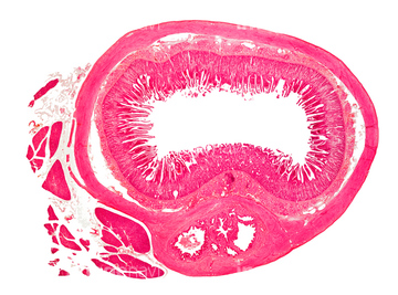
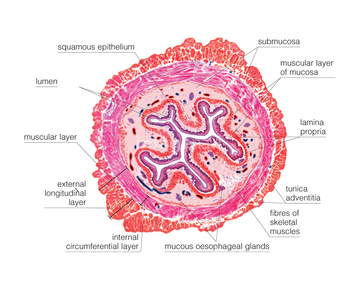
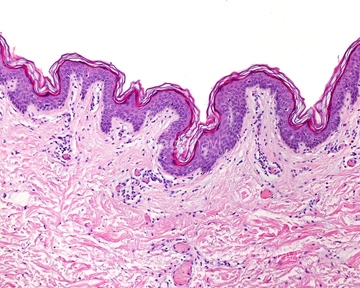
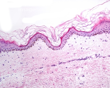
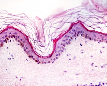
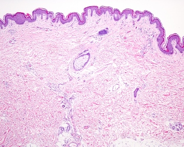
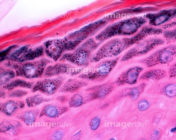
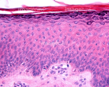
この検索結果には、Urinary bladder mucosa, light micrograph、Urinary bladder wall, light micrograph、Oesophagus, light micrograph、Stomach lining, light micrograph、Colon lining, light micrograph、Mackerel gills, light micrographなどが含まれています。

64146135

64146275
64221011
64206819
64066084

64234509
64234510
64196734
64073274
64073275
64073276
64235009
64235010
64114552
64114549
64114570
64114571
64224842
64073272
64073273
64008744
64008745
64063011
64063015
64197478
64071988
64071989
64084027
64234508
64114561
64177447
64225131
64197456
64008746
64008751
64093995
64065986
64088791
64076360
64076361
64221018
64225260
64213913
64213914
64213915
64085267
64114553
64225261

64225133

64234840
64234841
64234843

64225140
64073333

64235008
64251226
64251229
64235143
64235146
64235147
64235148
64225189
64114540
64114554
64114521
64224938
64072028

64072029
64098209
64196715
64085261
64106080

64225083
64234842
64225259
64061536
64190664
64206818
64197472
64224841
64225092
64167115
64167118
64180293
17202471
17202472
64225176
64114526
64114529
64190237
64114573
64114574
64161259
64161268
64207219
64207239
64207274
64066066
64066076
64085317
64085318
64136447
64071833
64071834
64167101
64167102
64167105
64167113
64167114
64167116
| 次ページ |
