HOME > 写真 > 科学・テクノロジー > 科学 > DNA・細胞
10,000件の写真素材が検索されました。
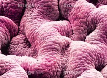
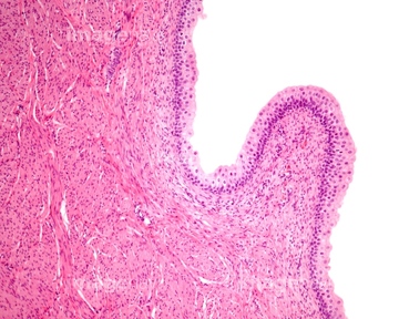
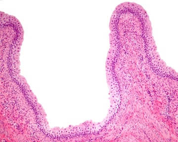
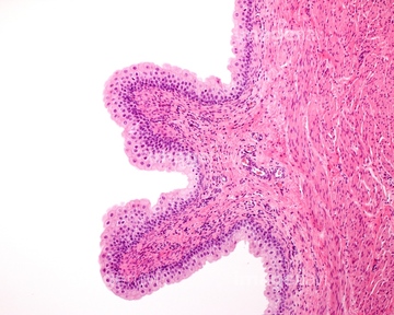
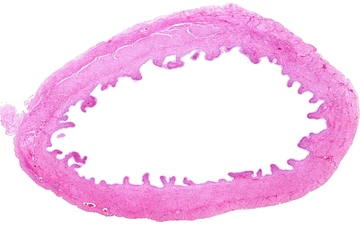
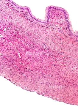
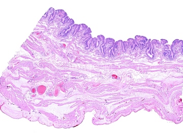
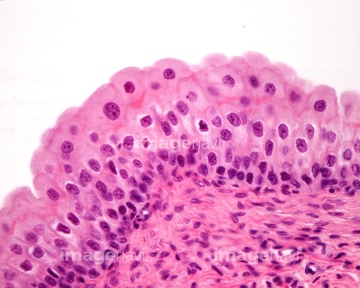
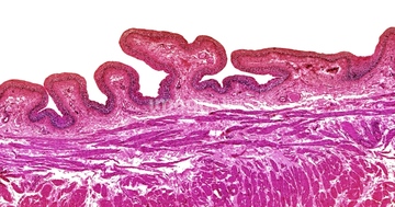
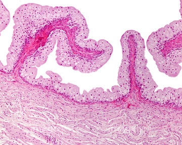
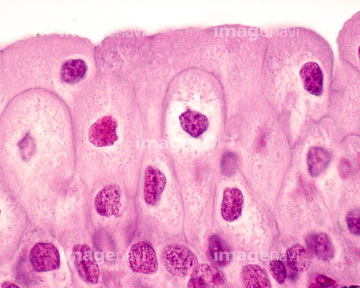
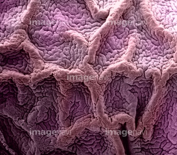
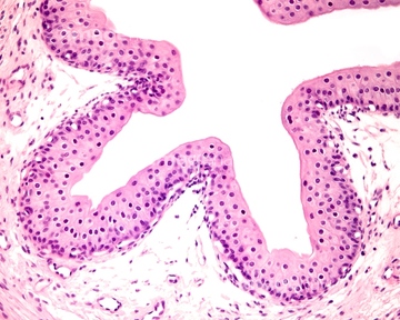
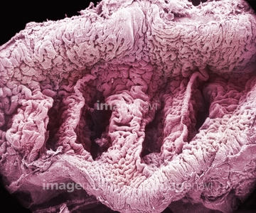
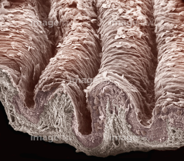
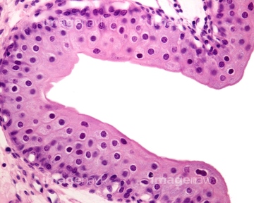
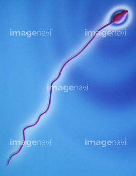
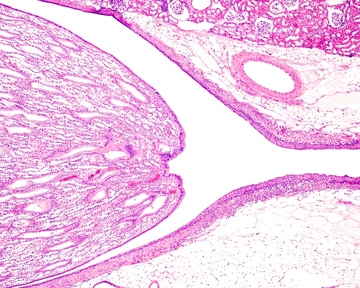
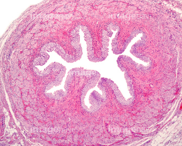
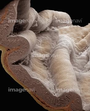
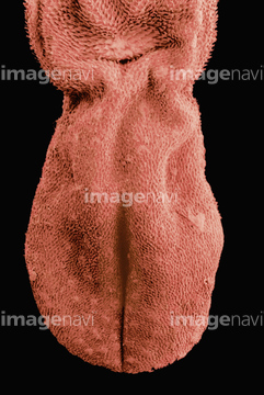
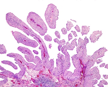
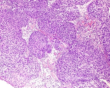
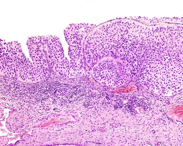
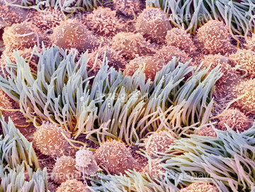
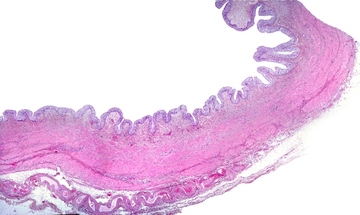
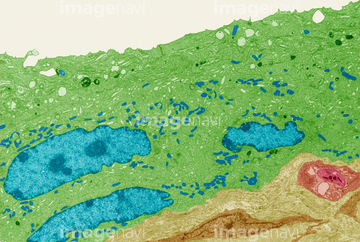
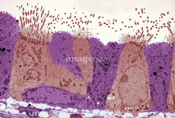
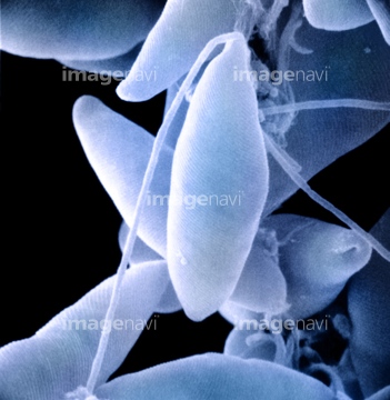
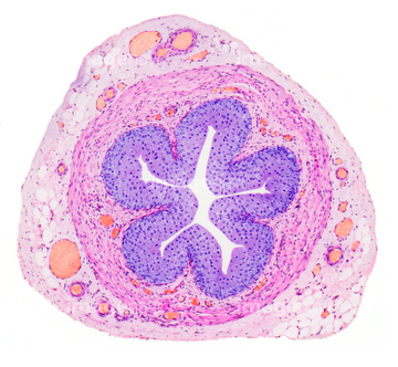
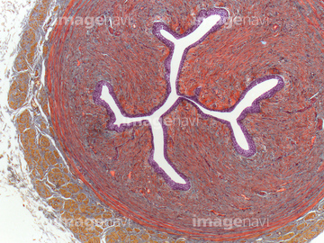
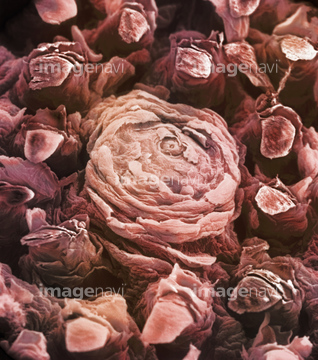
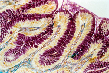
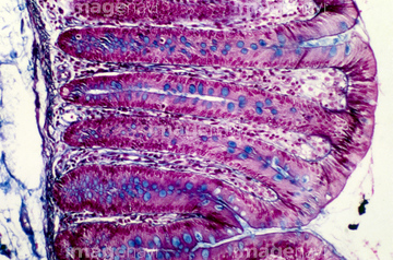
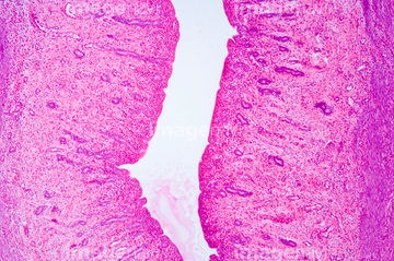
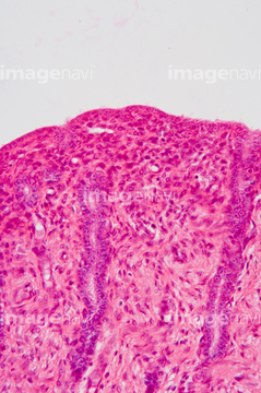
この検索結果には、Human sperm SEM X2250、Renal pelvis, light micrograph、Ureter, light micrograph、Stomach surface and wall section. SEM、Gallbladder, light micrograph、Gallbladder wall, light micrographなどが含まれています。
64225259
64197456
64254349
64098237
64245221
64225260
64190664
64225261
64206818
64197472
64207248
64207254

64221011
64225131

64225140
64225256
64115471
64115493
64115600
64225361
64205317
64245220

64146135

64146275


64225201
64085308
17281852
17281881
17281895
64234639
64234640
64207245
64114169

64225083

64225133
64225151
64225152
64254345
64152547
64225092
64225189
17246260
17246264
17246265
64115490
64115520
64115526
64115594
64115595
64224854
64225257
64114730
64224880
64225175
64225192
64115602

64104713
64104724
64104731
64104736
64056751
64146158
64060847
64115596
64225190
64161259
64161268
64008744
64008745
64219690
64219691
64219692
64219693
64225088
64225086
64225091
64225242
64225243
64225244
64225264
64093995
64088791
64225082
64136447
64206819
64235009
64235010
64196734
64197478
64066084
64071988
64071989
64114526
64114529
64114552
64037351
64225101

64055760
64083936
64115521
64225265
20528350
20528352
20528360
64103784
64103785
64225130
20523355
20523366
64045958
64190237
64008746
64008751
64115535
64115536
64114561
64203433
64245206
64245207
| 次ページ |
