HOME > 写真 > 科学・テクノロジー > 科学 > DNA・細胞
10,000件の写真素材が検索されました。
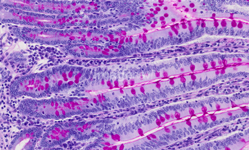
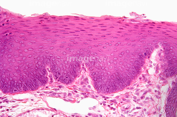
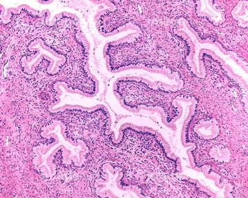
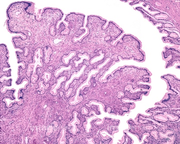
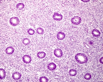
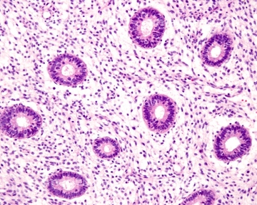
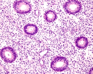
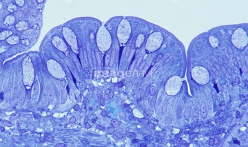
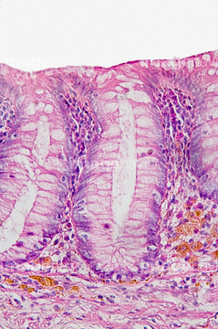
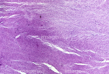
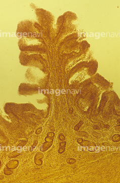
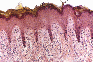
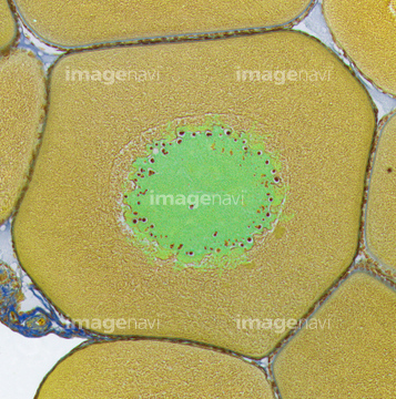
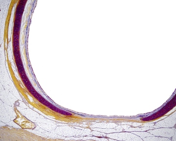
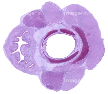
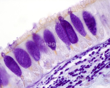
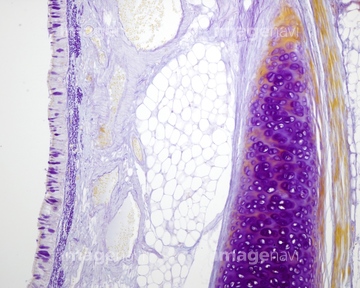
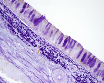
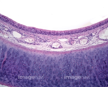
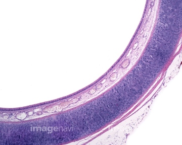
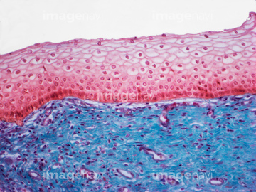
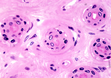
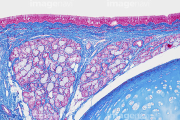
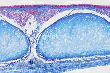
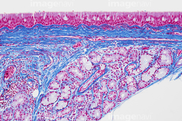
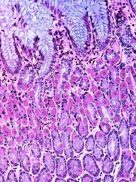
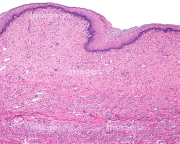
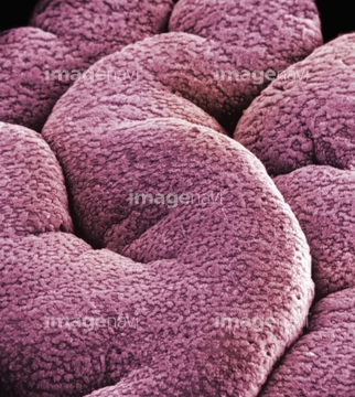
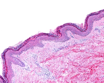
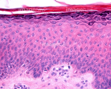
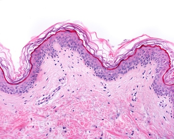
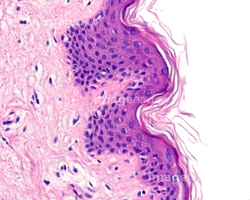
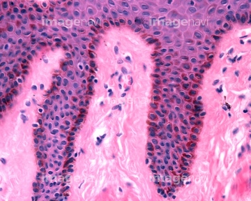
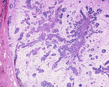
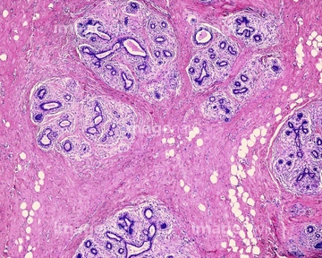
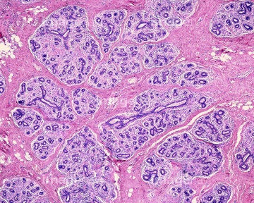
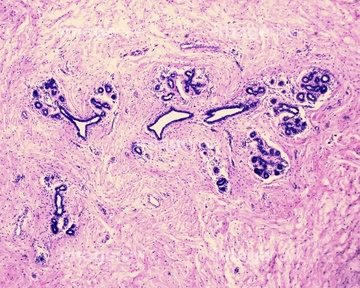
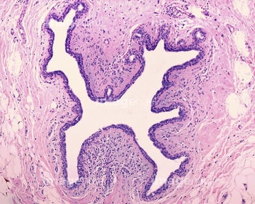
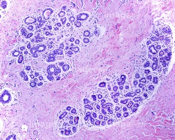
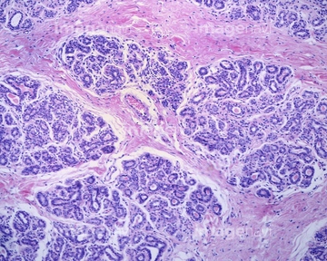
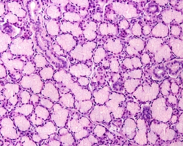
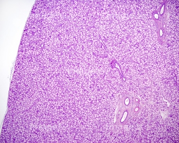
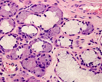
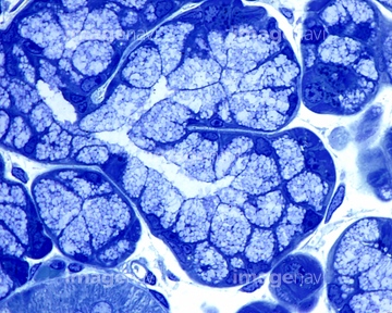
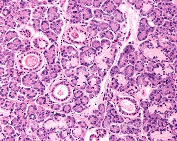
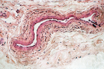
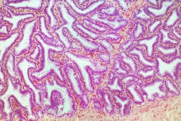
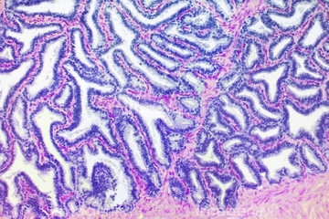
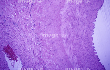
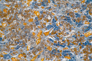
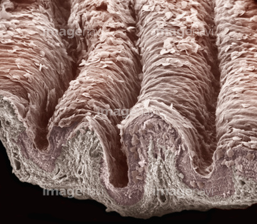
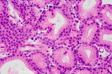
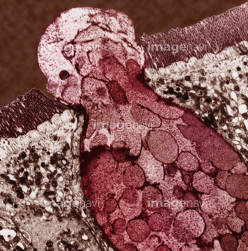
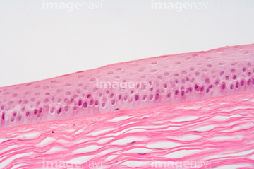
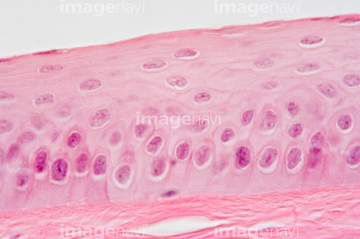
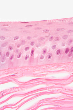
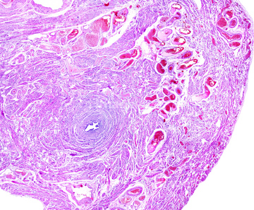
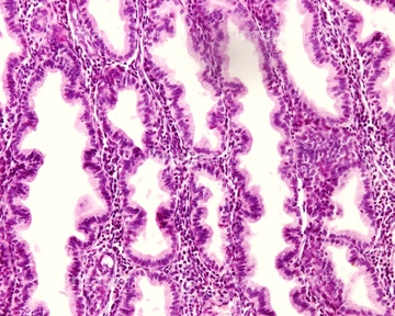
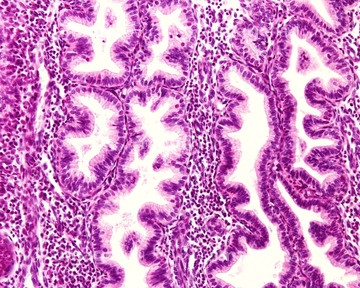
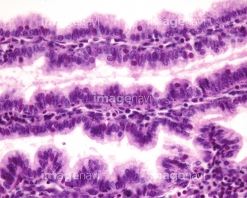
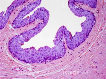
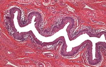
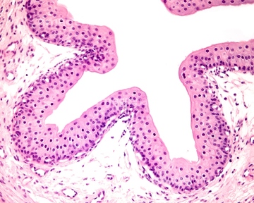
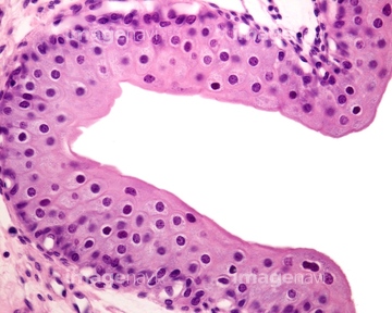
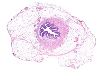
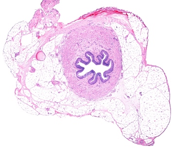
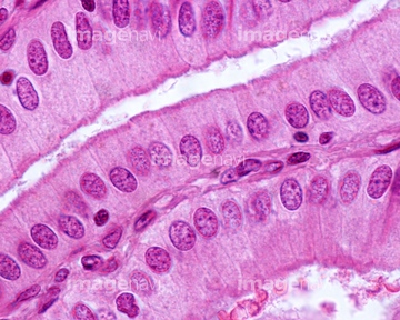
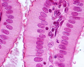
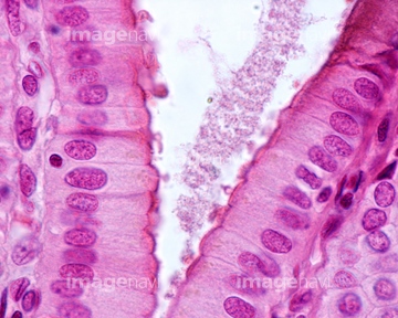
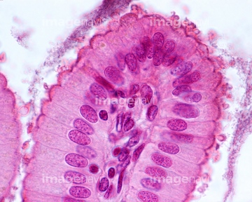
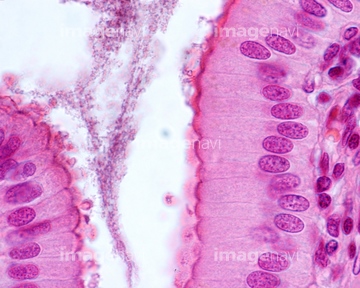
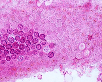
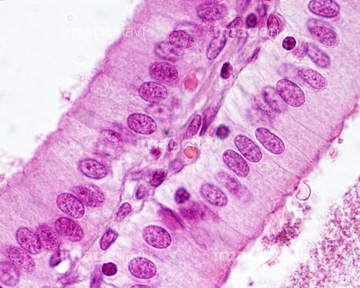
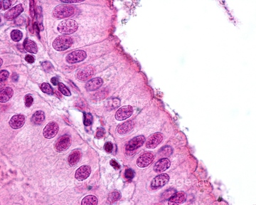
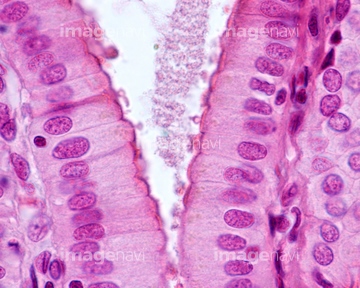
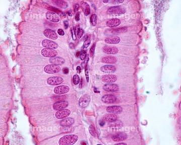
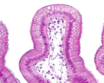
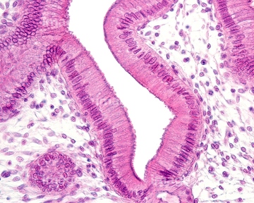
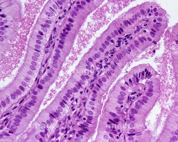
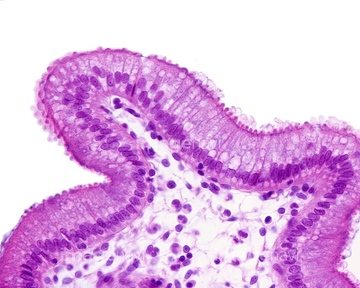
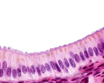
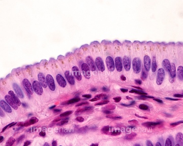
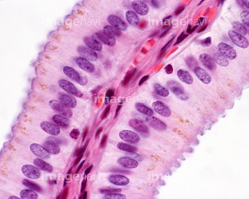
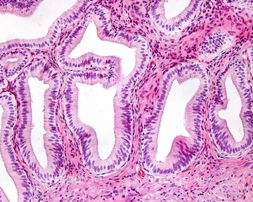
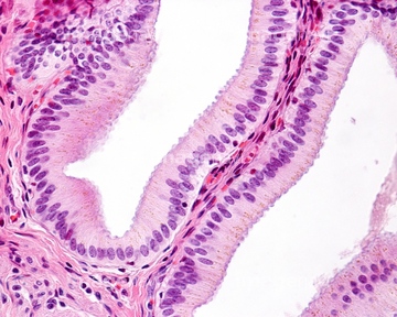
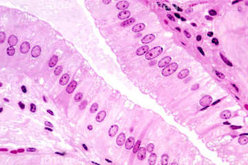
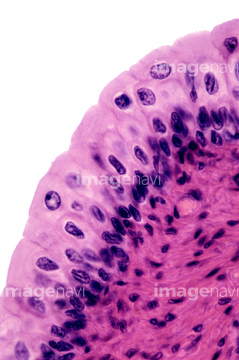
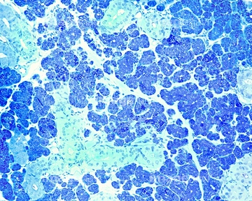
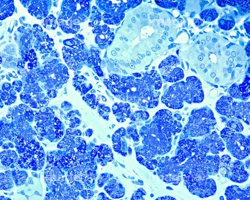
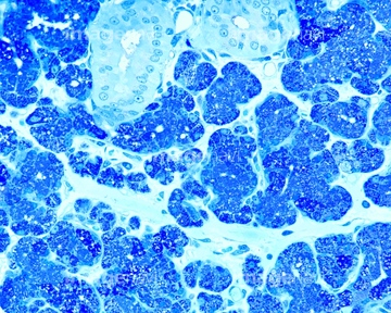
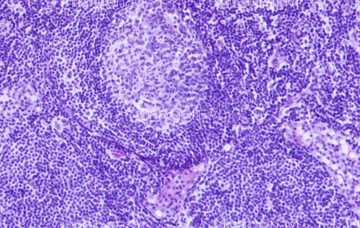
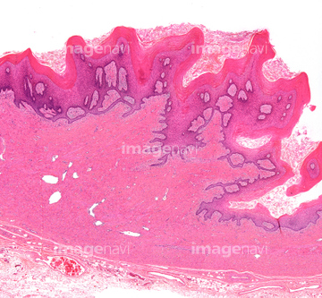
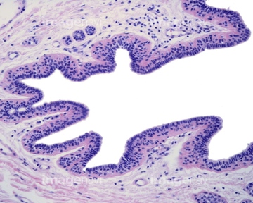
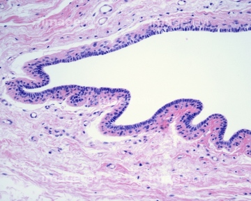
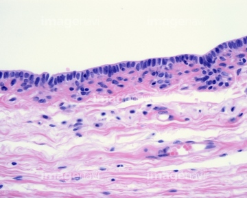
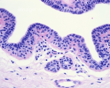
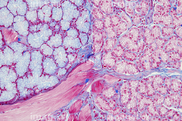
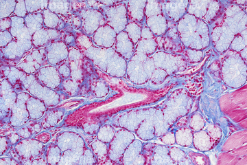
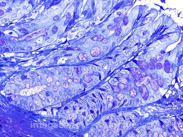
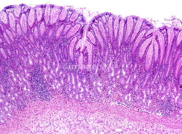
この検索結果には、Respiratory epithelium, light micrograph、Tracheal mucosa, light micrograph、Trachea, light micrograph、Human vagina, light micrograph、Human sweat glands LM X160、Gastric mucosa, light micrographなどが含まれています。
64114526
64114529
64114573
64114574
64251226
64251229
64114561

64177422
64209491
64209492
64209493
64116645
64114552
64207217
64207264
64234961
64224945
64198231
64198232
64234458
64234459

64146135

64146275
64207243
64207272
64207273
64197485

64114570
64114571
64114540
64205878
64205903
64088773
64088787

64161677
64161678
64161681
64161683
64161684
64093993
64224841
64235130
64235134
64235137
64251237
64167101
64167102
64167104
64167105
64167108
64167110
64167113
64167114
64167115
64167116
64167117
64167118
64167119
64167121
64167122
64167123
64167124
64167127
64167128
64180278
64180279
64180280
64180282
64180293
64090158
64106063
64177435
64146136
64146142
64146246
64146247
64146248
64146249

64115571
20548481
20548491
64115581
64225094
64114522
64114523
64114524
64114549
64114521
64177446
64225202
20532073
20532081
64234460
64234461
64116647
17256229
64169408
64169409
64190664
64206818
64197472
64221011
64224842
64224953
64090181
64180295
64180296
64180297
64180298
64177432
64177433
64177434
64177436
64177437
64225092
64235136
64235255
64235256
64090159
64090174
64090187
64098230
| 次ページ |
