HOME > 写真 > 科学・テクノロジー > 科学 > DNA・細胞
10,000件の写真素材が検索されました。
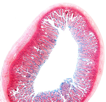
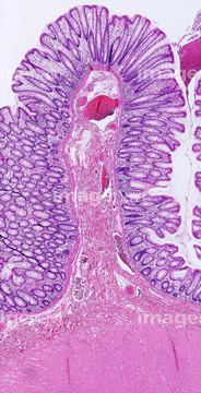
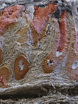
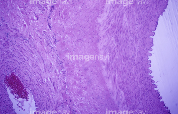
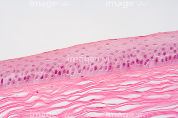
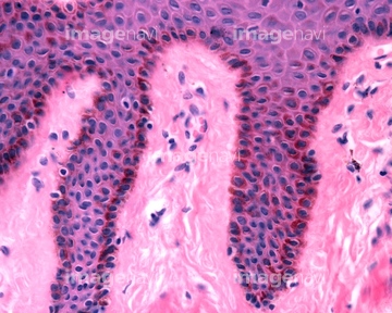
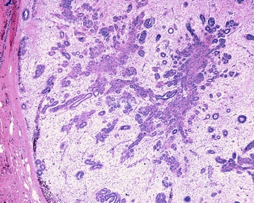
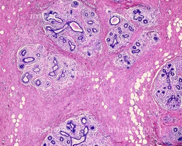
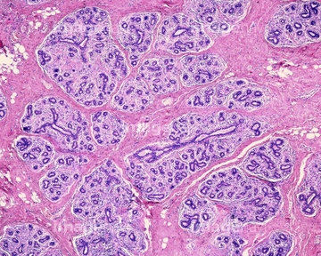
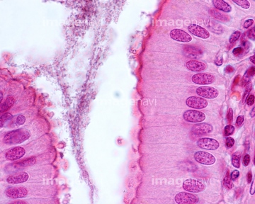
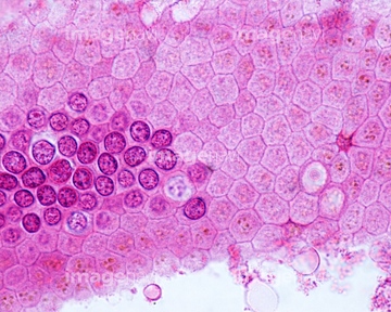
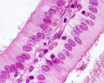
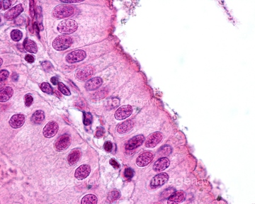
この検索結果には、Colon, SEM、Cross-section of the mammal colon. SEM、Section of an ulcer in the duodenum. LM、Small intestine goblet cells, light micrograph、Trachea, LM、Trachea, light micrographなどが含まれています。
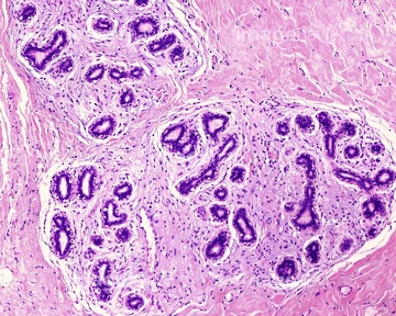
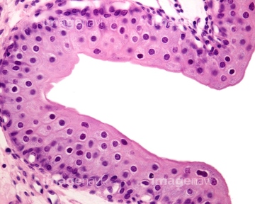
64114574
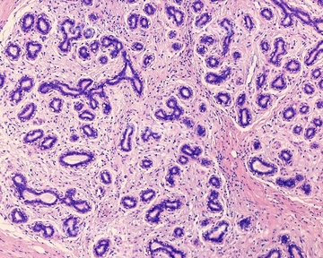
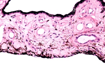
64114573
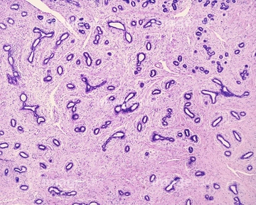
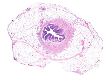
64114526
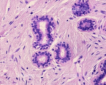
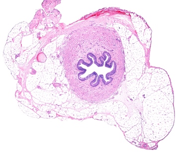
64114529
64090158
64106063
64251226
64251229
64234460
64234461
64090187
64225131
64090159
64116645
64207233
64207235
64071989
64114552

64251237
64177422

64146135

64146275
64114561
64066076
64197485
64225094
64161677
64161678
64161681
64161683
64161684
64090174
64224843
64114553
64114554
64088311
64088344
64088315
64250037
64250607
64250609
64250615
64207243
64207272
64207273
64235137
64116647
64167115
64167118
64180293

64088310
64224953
64090463
64162985
64114522
64114523
64114524
64114549
64114570
64114571

64225083

64225133
64235130
64235134
64167101
64167102
64167104
64167105
64167108
64167110
64167113
64167114
64167116
64167117
64167119
64167121
64167122
64167123
64167124
64167127
64167128
64180278
64180279
64180280
64180282
64180295
64180296
64180297
64180298
64150226
64150227
64146136
64146142
64146246
64146247
64146248
64146249

64098227
64254758
64090172
64073331
64190664
64190691
64206818
64197472
64221011
64224841
64224842
64213935
64072040
64073309
64073352
20542530
20544099
64214176
64057542
64085258
64073304
64073344
64225092

64098230
| 次ページ |
