HOME > 写真 > 科学・テクノロジー > 科学 > DNA・細胞
10,000件の写真素材が検索されました。
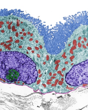
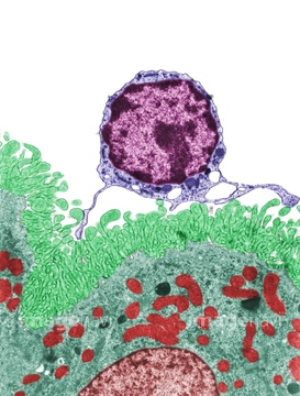
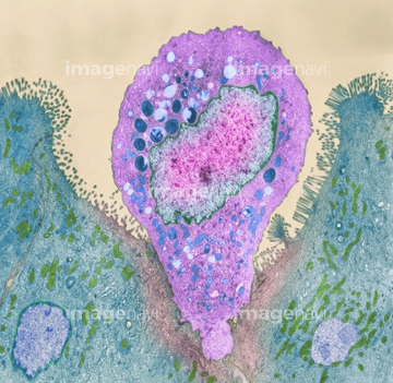
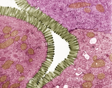
この検索結果には、Hairy scalp skin, light micrograph、Intestinal gland cell, TEM、Endocrine cell, TEM、Intestinal microvilli, SEM、LM of an intestinal villus in cross section、Small intestine villi, light micrographなどが含まれています。
64056269
64008073
64008776
64247699
64060201
64060202
64166169
64247715
64247717
64060847
64055781
64259990
64061536
64075661
64209800
64056271
64062279
64055763
64077441
64062297
64062298
64225131
64251226

64225083

64225133
64049910
64049911
64190895
64057593
64057594
64057595
64057596
64057597
64057598
64062293
64062294
64063628
64063629
64063630
64063631
64042017
64042018
64065626
64103901
64064550
64060814

64060815
64060816
64022357
64022358
64060825
64060826
64196734
64206819
64251229
64066076
64071989
64167311
64265564

64265567
64224843
64114169
64077392
64078784
64078785
64082457
64076318
64076319
64076358

64076359
64076360
64076361

64076371
64076372
64225151
64225152
64056944
64057590
64057591
64057592
64061547
64045916
64074874
64075100
64076019
64150280
64068337
64223998
64223999
64029680
64029681
64029682
64145944
64145945
64169972
64152547
64209759
64264588
64264589
64209773
64209785
64209786
64211723
64211724
64211725
64247698
64060808
64060809
64060810
64063632
64063633
64190892
64190893
64190894
64147797


64104713
64104724
64104731
64104736
20548481
20548491
64059680
64089586
64089593
64225092
64040543
64040544
64147753
64132269
64132270
64060827
64060828
64265553
64265570
64265573

64146135

64146275
64114552
64212991
64213007
64105465
64105477
64021603
64021607
64105462
64105475
64183427
64183428
64183429
64183430
64244240
64247694
64247695
64247697
64247705
64247710
64161259
64161267
64161268
64161269
64161281
64098237
| 次ページ |
