HOME > 写真 > 人物 > 構図 > 色バック
10,000件の写真素材が検索されました。
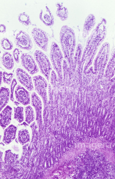
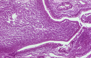
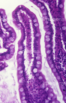
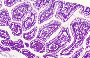
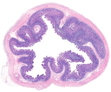
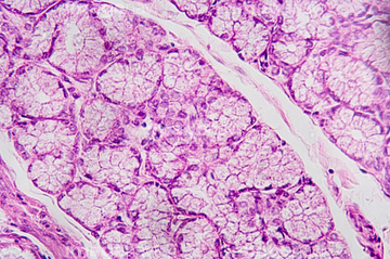
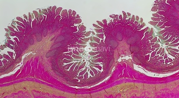
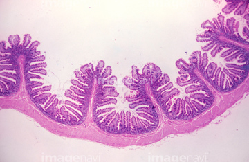
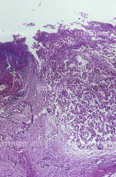
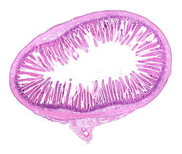
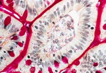
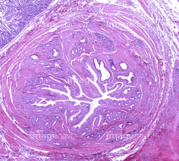
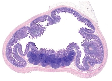
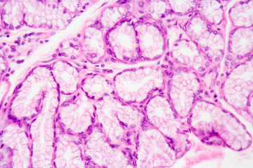
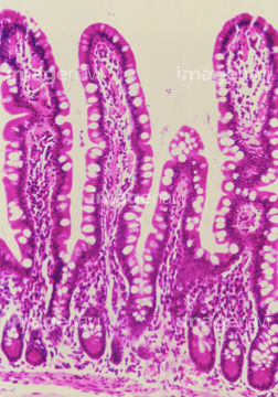
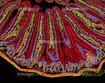
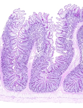
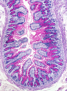
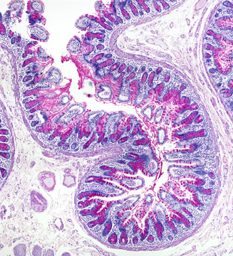
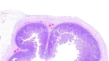
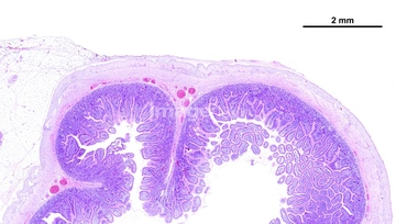
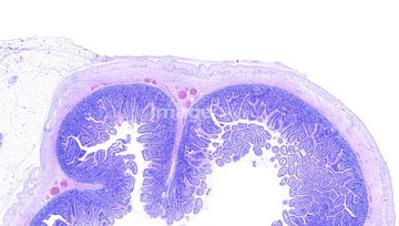
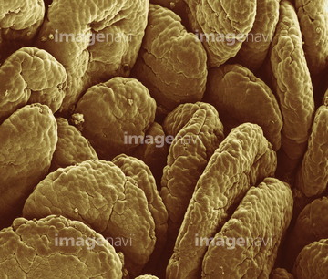
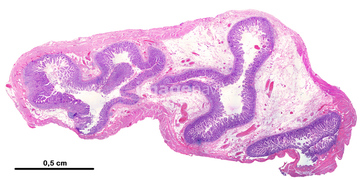
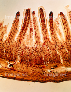
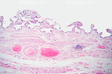
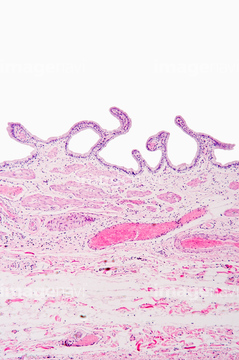
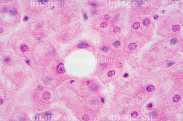
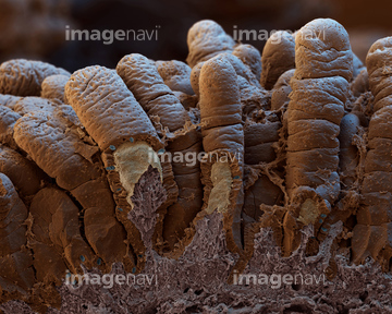
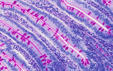
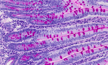
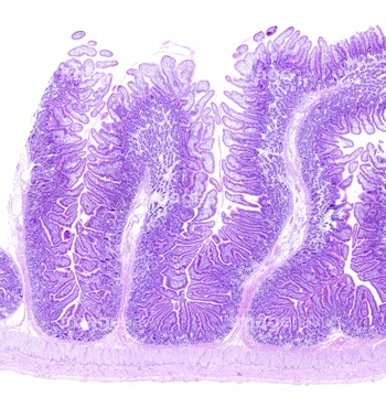
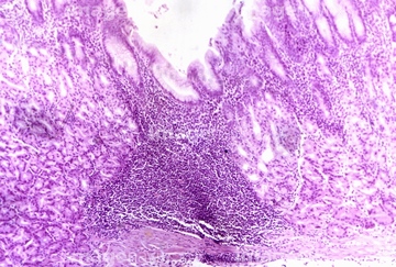
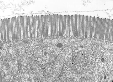
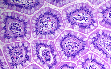
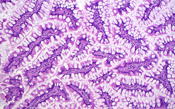
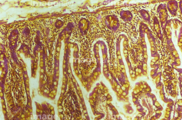
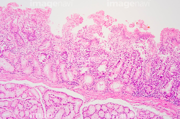
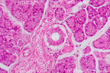
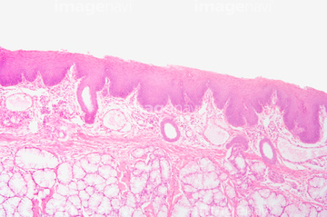
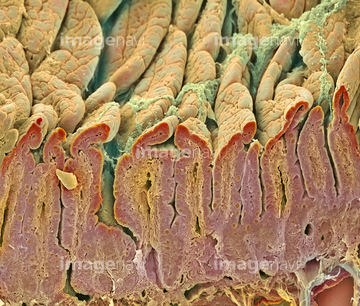
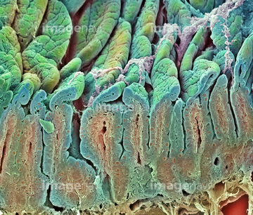
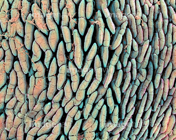
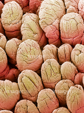
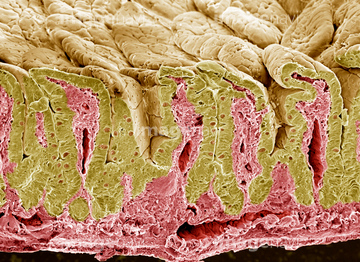
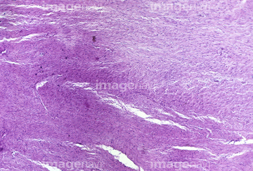
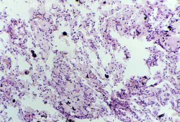
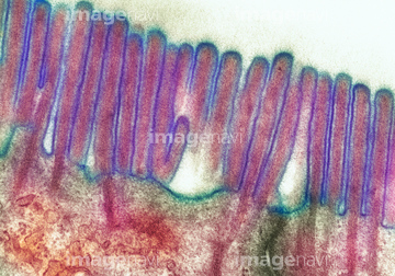
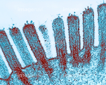
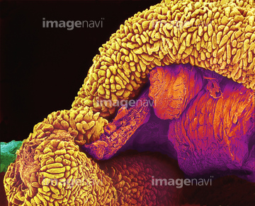
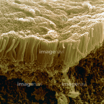
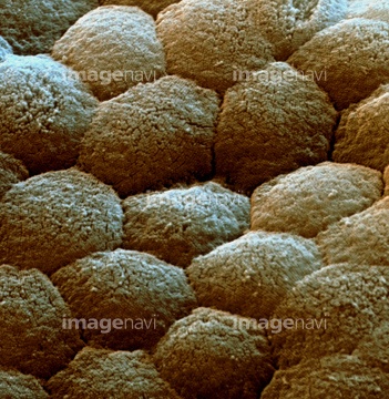
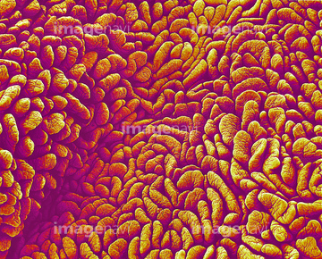
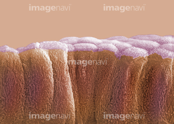
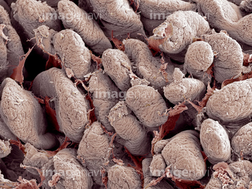
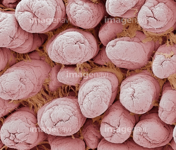
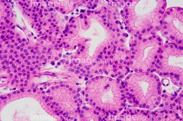
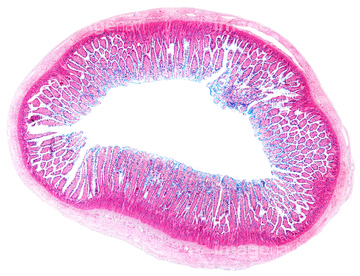
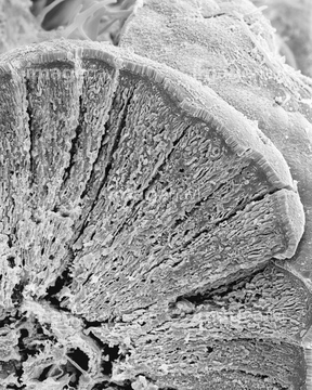
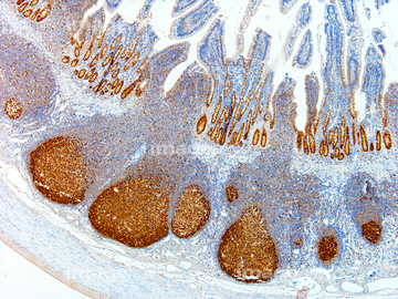
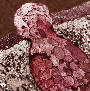
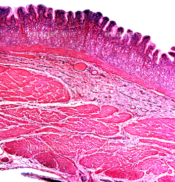
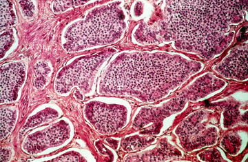
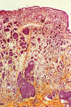
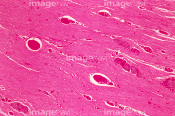
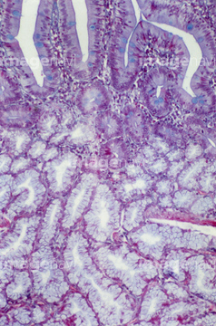
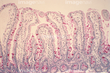
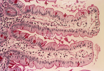
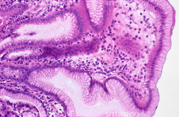
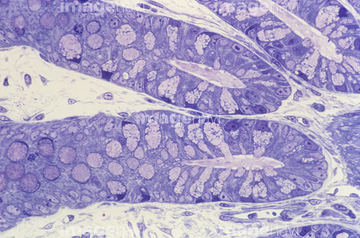
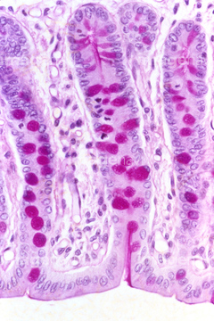
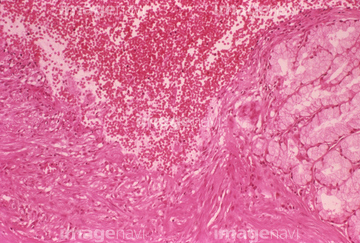
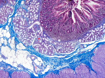
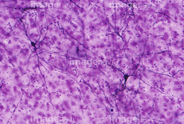
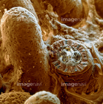
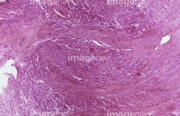
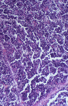
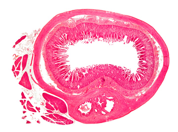
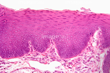
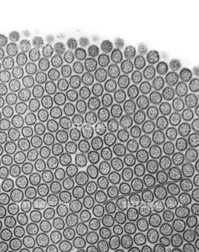
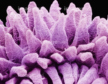
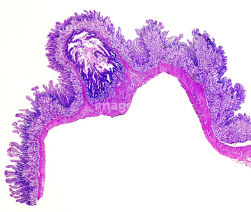
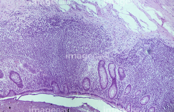
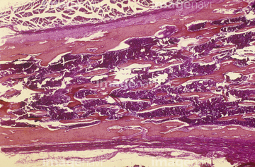
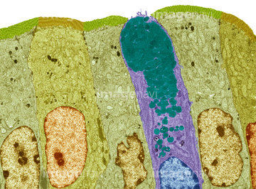
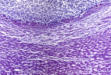
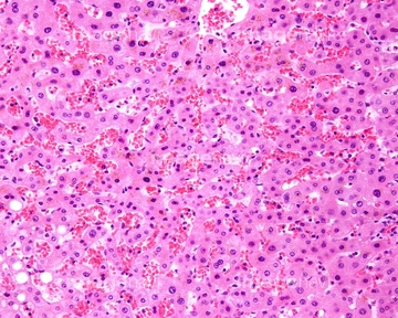
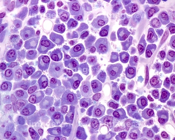
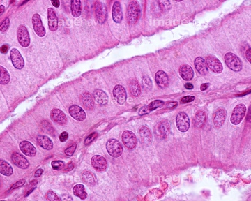
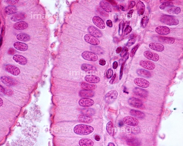
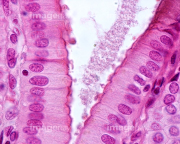
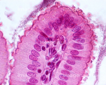
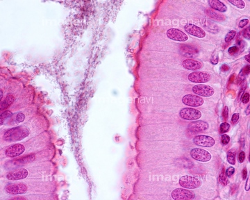
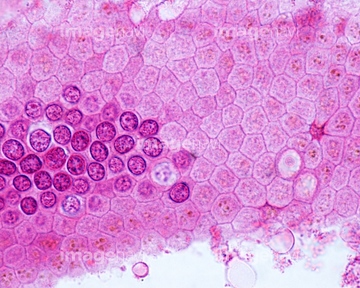
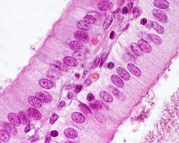
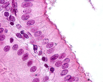
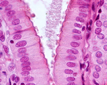
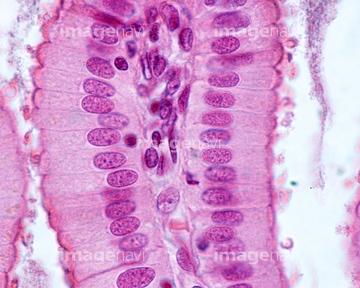
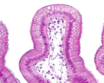
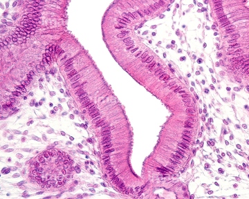
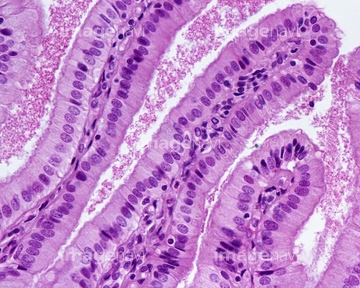
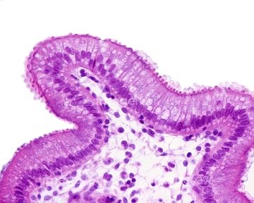
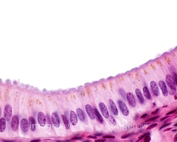
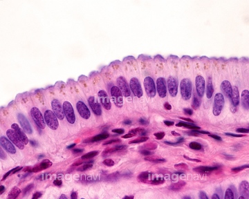
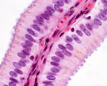
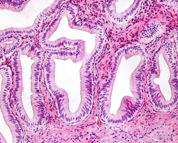
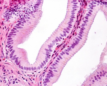
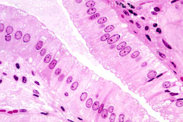
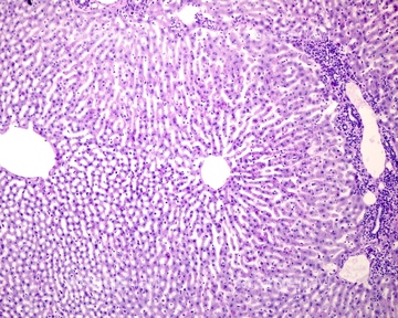
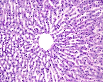
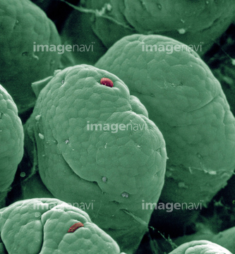
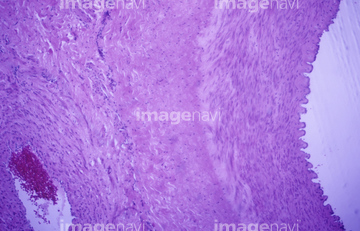
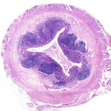
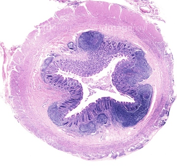
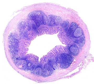
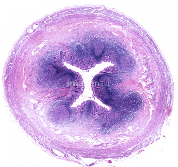
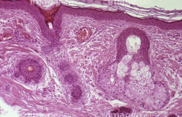
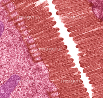
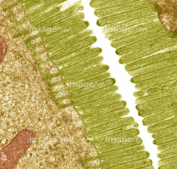
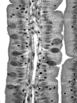
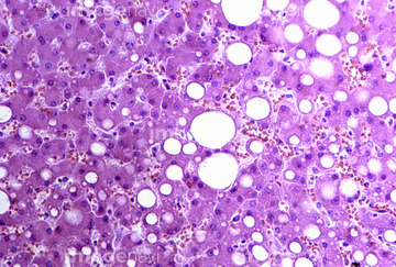
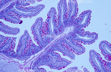
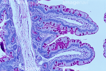
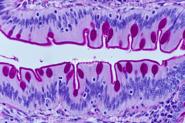
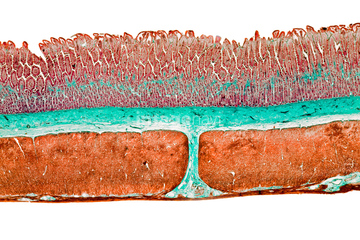
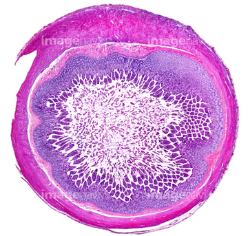
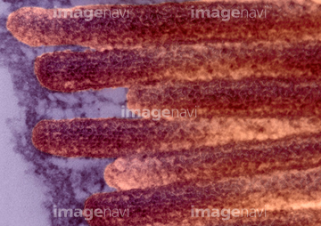
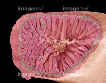
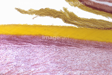
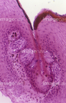
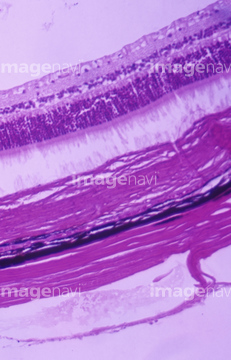
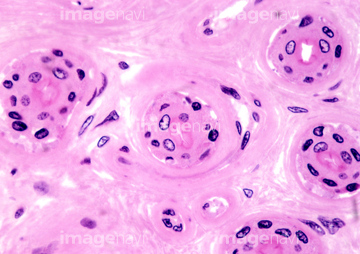
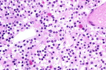
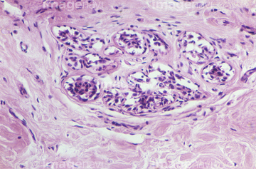
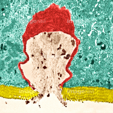
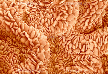
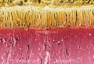
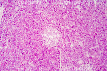
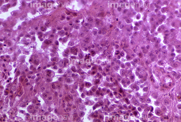
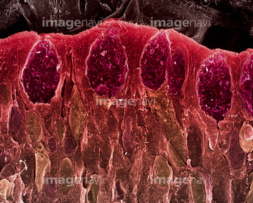
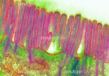
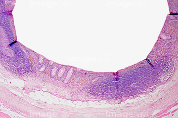
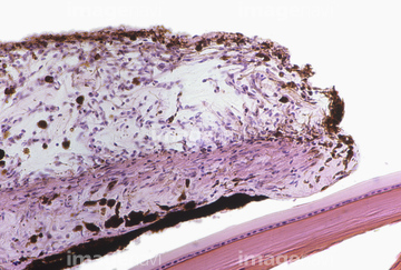
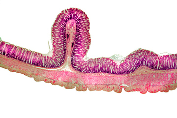
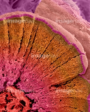
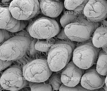
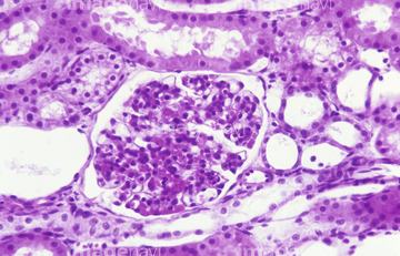
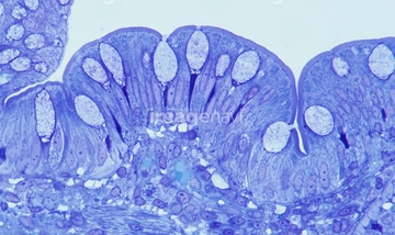
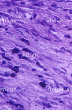
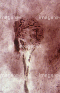
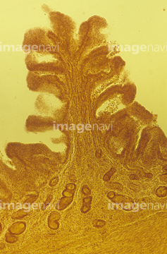
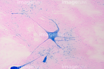
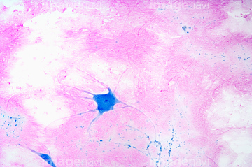
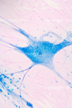
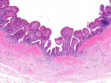
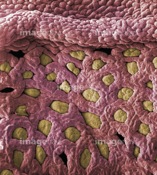
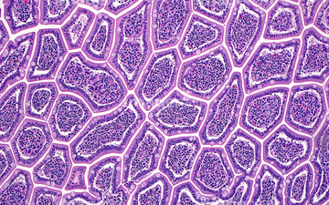
この検索結果には、Intestinal lining, SEM、Human uterus section showing fibroids. LM、Collapsed human lung tissue LM X140、Duodenal microvilli、Intestinal microvilli, TEM、Villus surface of the small intestine, SEMなどが含まれています。
64224937
64224938

64225083

64225133
64197487
64114553
64196715
64197488
64114554
64021570
64206819
64090479
64090480
64213913
64213914
64213915
64021569
64114570
64114571

64225140
64114521
64021534
64251226
64251229
64196734

64225245
64247696
64256459
64114549
64021539
64021540
64021541
64021568
64021571
64224932
64021528
64021545
64088343
64114540
64114552
64021532
64021535
64021536
64021538
64225176
64008180
64075661
64090152
64225094
64072010
64072070
64072071
64073343
64090490
64021533

64146135

64146275
64114561
64225256
64197106
64224942
64056271
64224904
64199179
64208212
64221011
64224842
64213931
64213933
64049910
64196724
64196725
64172510
64172534
64224950
64225224
64115581
64224951
17202471
17202472
64155048
64224907
64021549
64021550
64021551
64197097
64055781
64061536
64224841
64224844
64021554
64073608
64077368

64224908
64021537
64056746
64114551
64085258
64225203
64190181
64190182
64190183
64190184
64114577
64152547
64224954
64116645

64098209
64263188
| 次ページ |
