HOME > 写真 > 科学・テクノロジー > 科学 > DNA・細胞
10,000件の写真素材が検索されました。
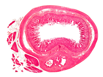
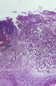
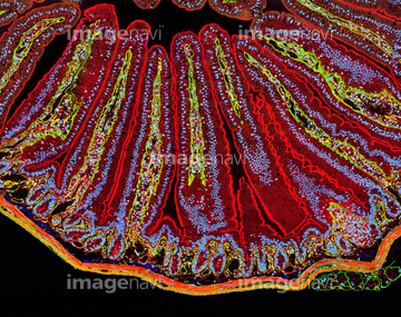
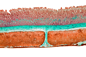
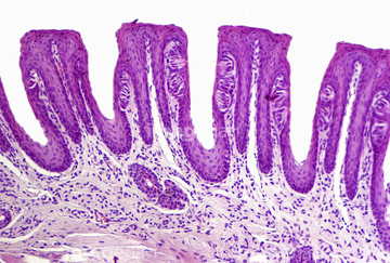
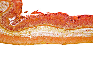
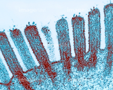
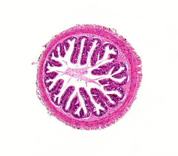
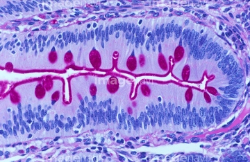
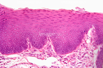
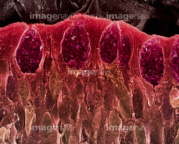
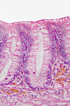
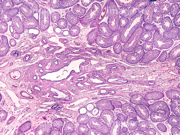
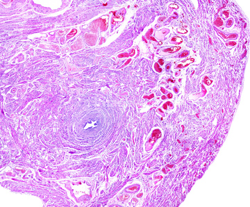
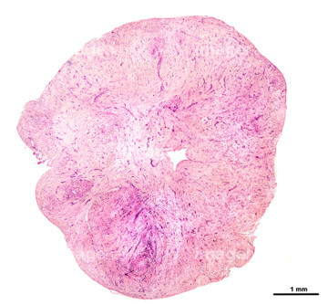
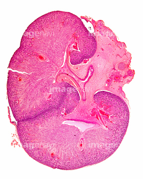
この検索結果には、Appendix, light micrograph、Oesophagus, light micrograph、LM of blood vessels in villi of small intestine、Cat colon, light micrograph、Villi in th small intestine、Cross-section of the human esophagus. LMなどが含まれています。
64196734
64224937

64225083
64251229
64196715
64206819
64251226

64225133
64225094
64224938

64225140
64021570
64234476
64234477
64234478
64234479
64116645
64060933
64234667
64234718
64021534
64114553
64114554
64213913
64213914
64213915
64196724
64196725
64172510
64172534
64021569
64073333
64085267
64085318
64085258
64225256

64021536
64090479
64090480
64224843
64083975
64060202
64021533
64072067
64021539
64021540
64021541
64021571
64085261
64021545
64204996
64116647
64114561
64021537
64114552
64225245
64073310
64088343
64147742
64114540
64114570
64114571
64075661
64049910
64021532
64021535
64021538
64114521
64247696
64234473
64234474
64073343
64172526
64021549
64021550
64021551
17202471
17202472
64056271
64150338
64073309
64196735

64146135

64146275
64021568
64055781
64254328
64172509
64085264
64085268
64085272
64085308
64254295

64021528
64021552
64221011
64224842
64021548
64152547
64114549
64197106
64225224
64021580
64256459
64008180
64066076
64224932
64073301
64132948
64132949
64225176
64090490
64197487
64197488
64072010
64072070
64072071
64061536
64049911
64021554
64077368
64055695
| 次ページ |
