HOME > 写真 > 科学・テクノロジー > 科学 > DNA・細胞
10,000件の写真素材が検索されました。
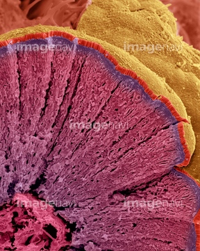
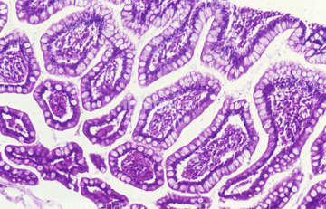
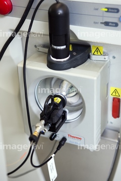
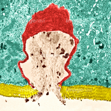
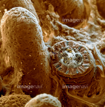
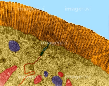
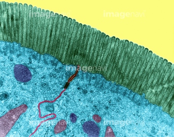
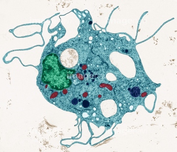
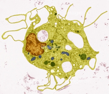
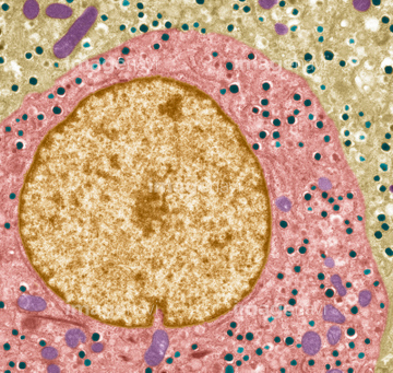
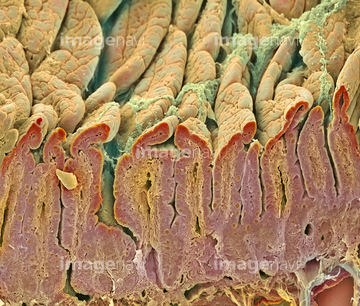
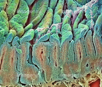
この検索結果には、Villi of the human small intestine. LM、Mass spectrometry injection mechanism、Goblet cell、Small intestine villi, SEM、Automated testing for bowel cancer、Choroid plexus epithelium, TEMなどが含まれています。
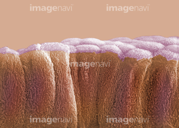
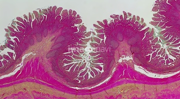
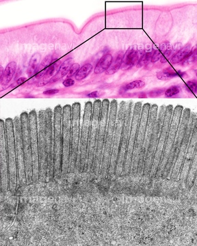
64225093
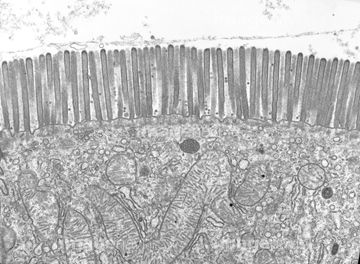
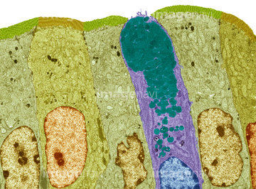
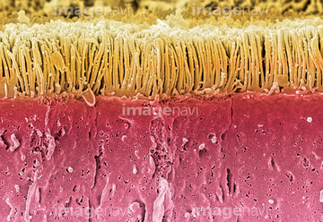
64225094
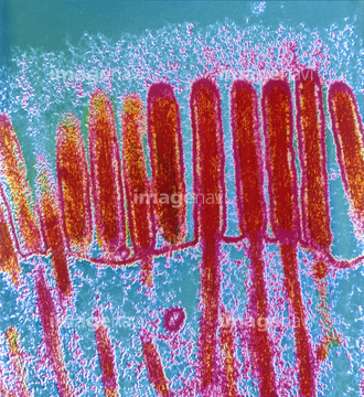
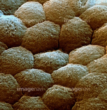
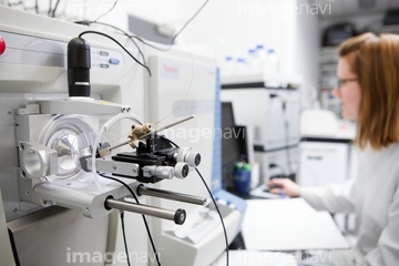
64021528
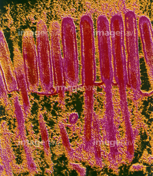
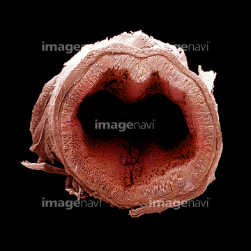
64021545
64225176
64247696
64264588
64264589

64225133
64021535

64225083
64014601
64014602
64179087
64179088
64224937
17202471
17202472
64055781
64190895
64204536
64204543
64204544
64056271
64021536
64115597
64060825
64060826
64060827
64060828
64056746
64209800
64021532
64021534
64021538
64225106
64190892
64190893
64190894
64021554
64021533
64049788
64223998
64223999
64055763

64146135

64146275
64114521
64114549
64114552
64114553
64114570
64114571
64224938
64133039
64199179
64208212
64221011
64224842
64213931
64213933
64021621

64225140
64103779
64103786
64021593
64021596
64021597
64021598
64021604
64021539
64021540
64021541
64021568
64021569
64021571
64021591
64021592
64114169
64225088
64225132
64105462
64105475
64114732
64114733
64088343
64147742
64145925
64060201
64060202
64166159
64166182
64077368
64225131
64114540
64114554
64225245
64190181
64190182
64190183
64190184
64204230
64204232
64209749
64209762
64209763
64209791
64209813
64021570
64196715
64206819
64090479
64090480
64213913
64213914
64213915
64203396
64203413
64203445
18597771
64245075
64225129
64225151
64225152
64225201
64115471
64244244
64029976
64224879
64103776
64103794
64167309
64167310
64209773
64209785
64209786
64211723
64211724
64211725
64225184
64002984
64002985
| 次ページ |
