HOME > 写真 > 科学・テクノロジー > 科学 > DNA・細胞
10,000件の写真素材が検索されました。
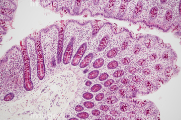
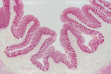
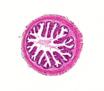
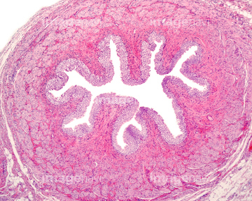
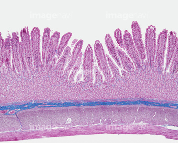
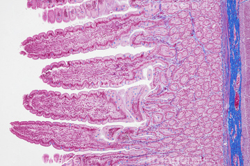
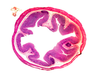
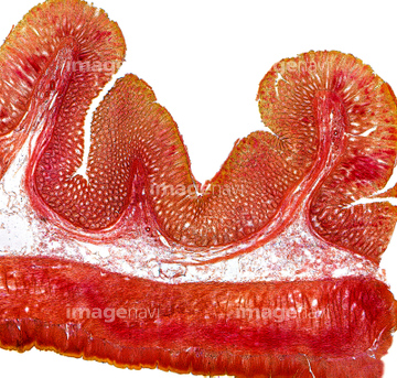
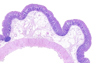
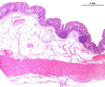
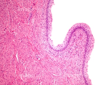
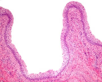
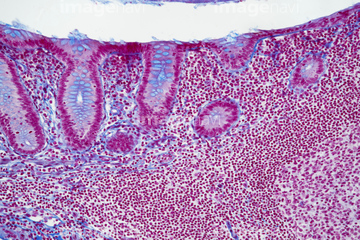
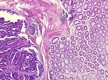
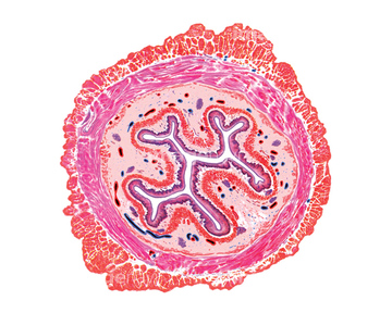
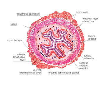
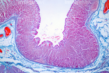
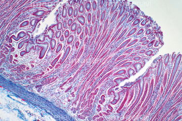
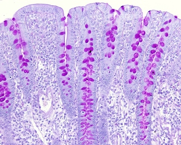
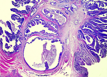
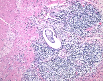
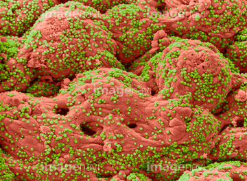
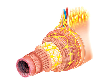
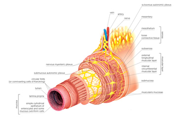
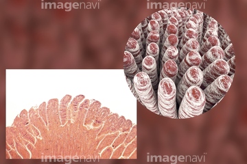
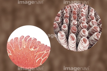
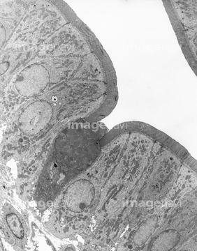
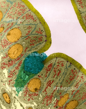
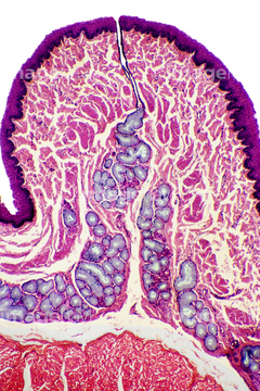
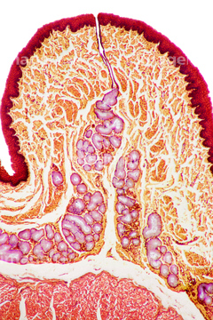
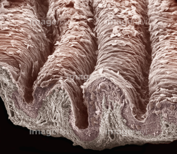
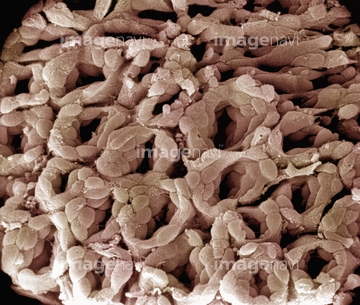
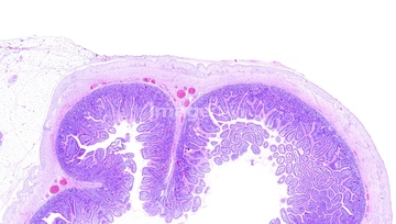
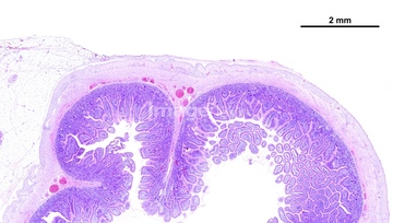
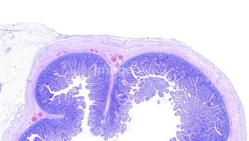
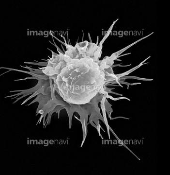
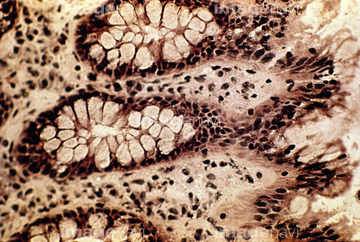
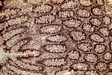
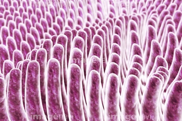
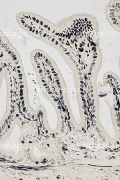
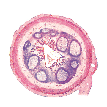
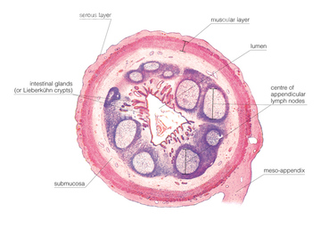
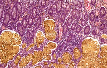
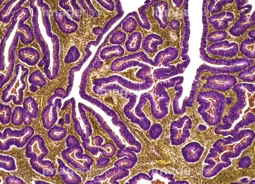
この検索結果には、Colon, LM、Appendix intestinal smooth muscle layers, light mi…、Large intestine, light micrograph、Large bowel, light micrograph、Urinary bladder mucosa, light micrograph、Urinary bladder wall, light micrographなどが含まれています。
64090172
64225131
64225192

64225140
64250037
64225256
64234460
64234461
64204996
64245220
64234471
64234472
64055695
64055694
64055754
64071892
64071893
64071989
64084027
64197486
64197489
64071876
64071877
64071886
64071887
64225260
64234476
64234477
64234478
64234479
64234667
64234718
64073272
64073273
64073274
64073275
64073276
64195478
64008746
64008751
64207233
64207235
64196735

64206746
64206747
64234838
64055692
64055693
64090158
64090159
64090463
64106063
64234280
64071833
64071834
64071850
64071851
20542530
20544099
64199159
64196498
64018223
64234469
64002980
17251192
17251193
17251194
64085258
64192928
64021523
64021524
64225259
64225261
64065178
64197140
64225095
64225242
64032267
64029976
64090458
64071988
64205008
64196715
64213913
64213914
64213915
64150240
64225265
64150338
64088311
64088344
17255816
64008159

64194255
64194256
64194257
64192929
64192930
64192931
64192932
64075581
64234508
64234509
64234510
64071902
64071903
64155646
64155653
64155666
64155671
64008754
64008755
64012176
64075583
64245080
64245081
64245082
64245083

64245084
64245087
18597803
17234583
21554723
21554727

| 次ページ |
