HOME > 写真 > 科学・テクノロジー > 科学 > DNA・細胞
10,000件の写真素材が検索されました。
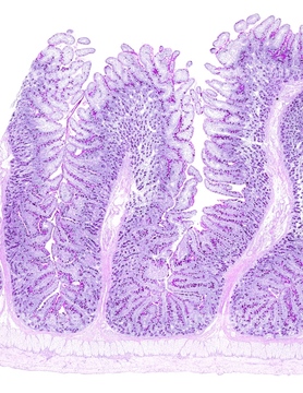
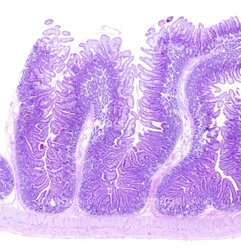
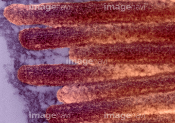
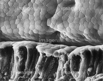
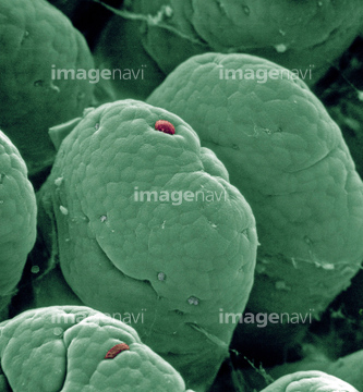
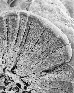
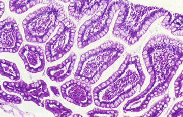
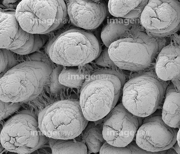
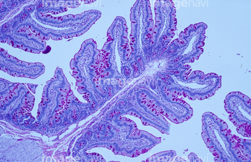
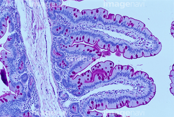
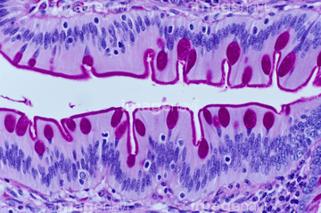
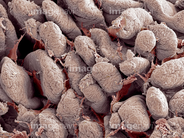
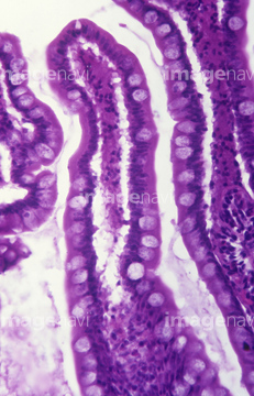
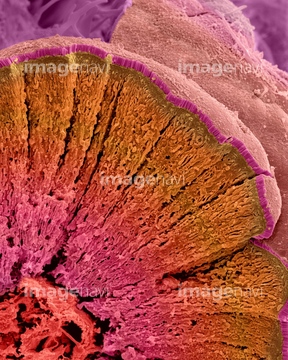
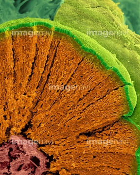
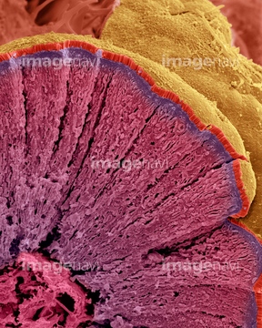
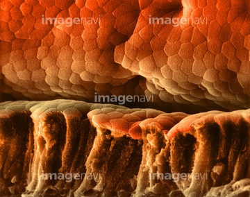
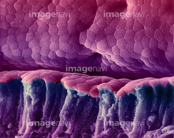
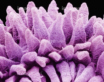
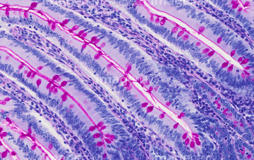
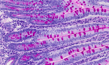
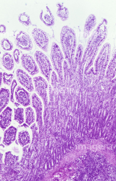
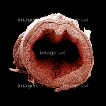
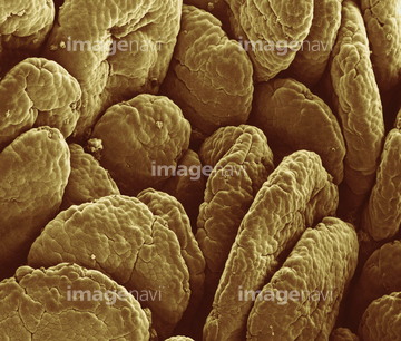
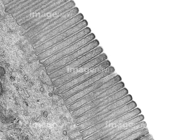
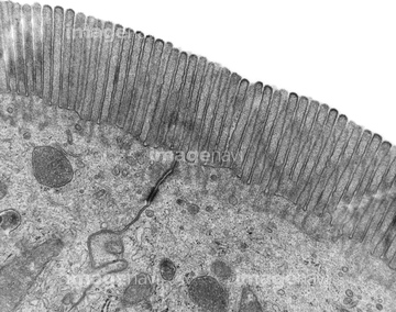
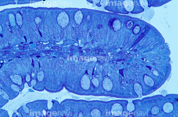
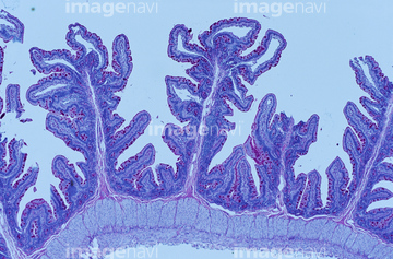
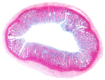
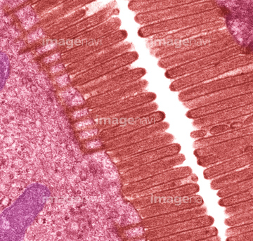
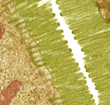
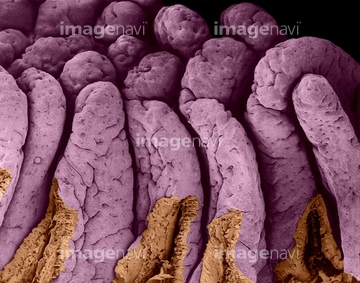
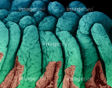
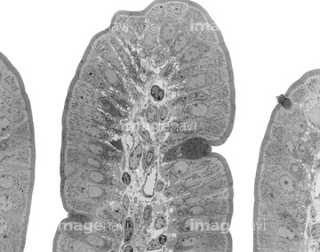
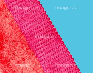
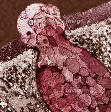
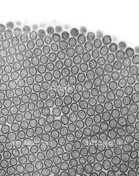
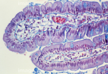
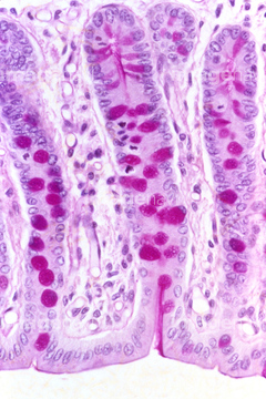
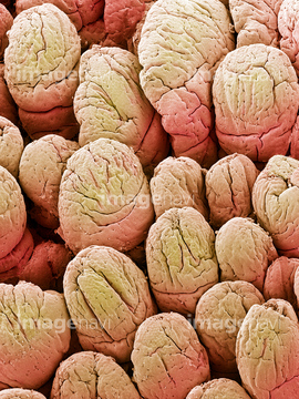
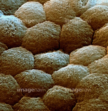
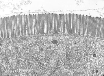
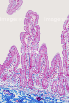
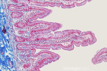
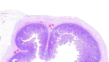
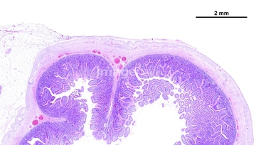
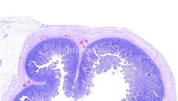
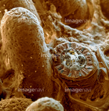
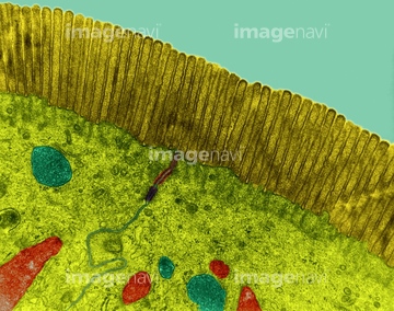
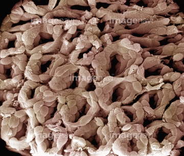
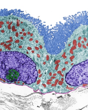
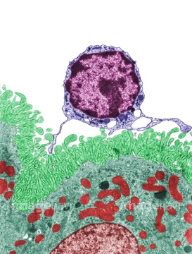
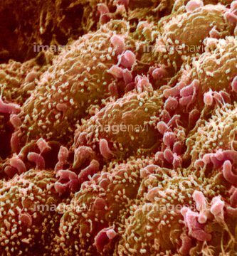
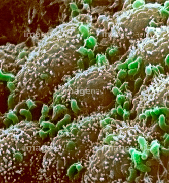
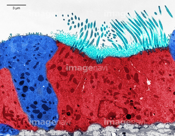
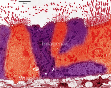
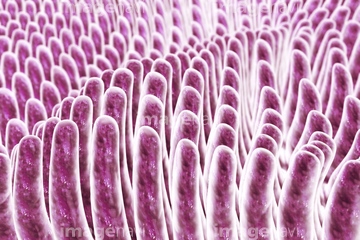
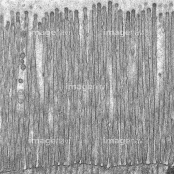
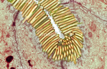
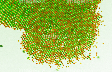
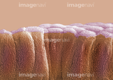
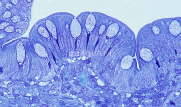
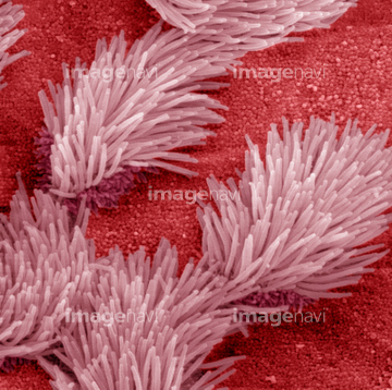
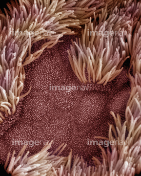
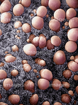
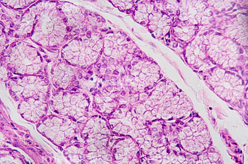
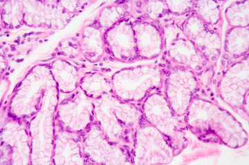
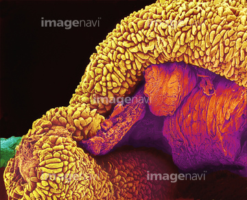
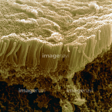
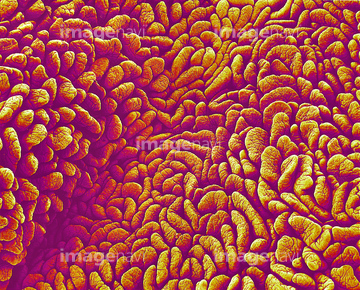
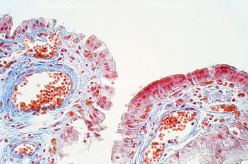
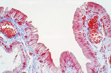
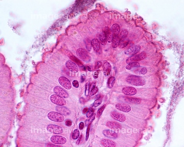
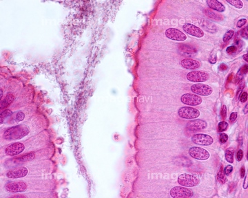
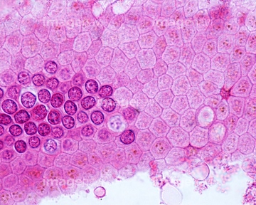
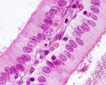
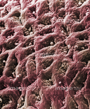
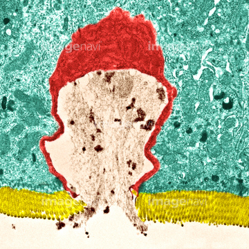
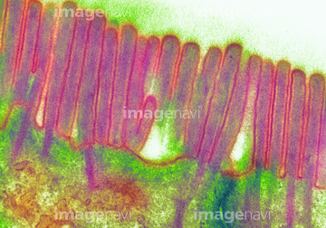
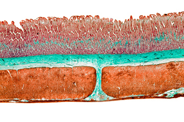
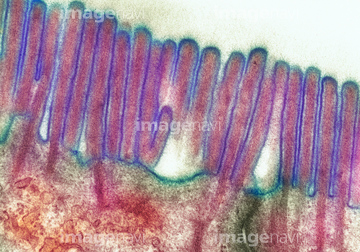
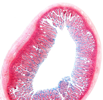
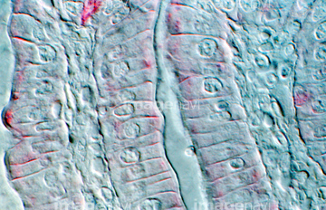
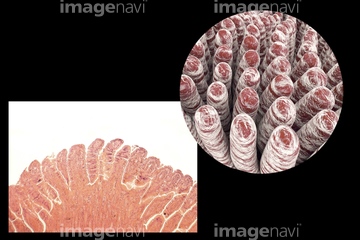
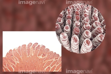
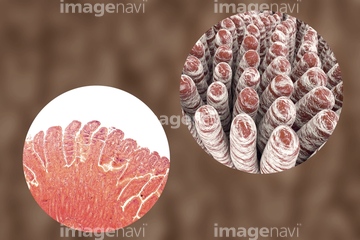
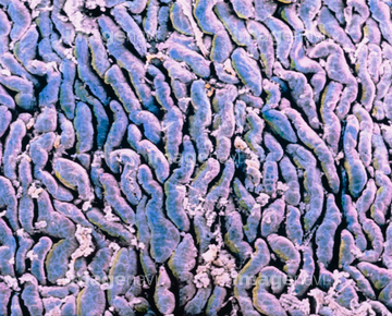
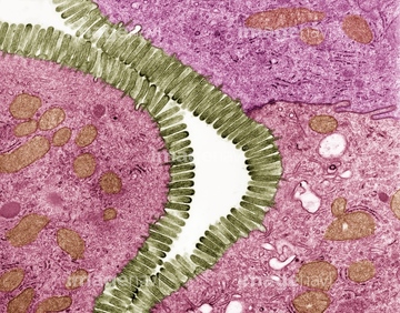
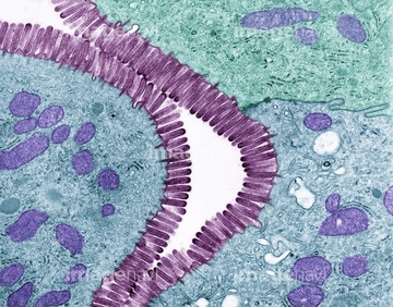
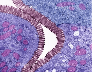
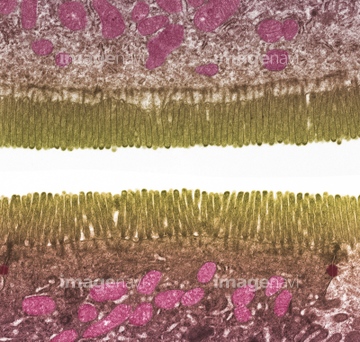
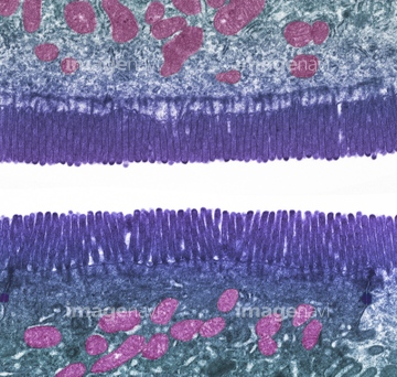
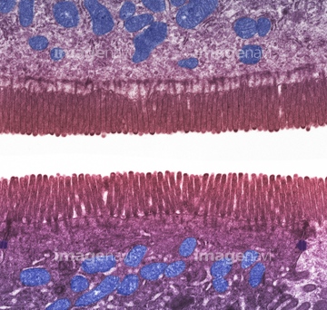
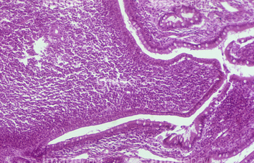
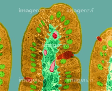
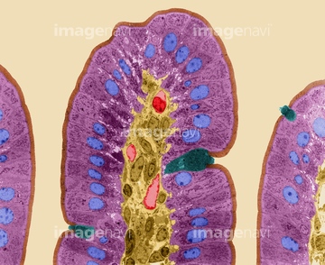
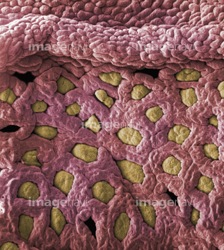
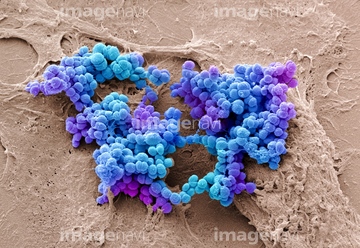
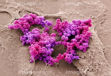
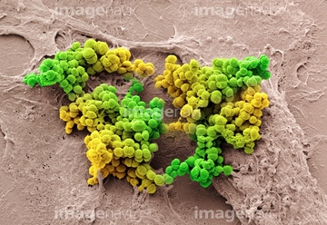
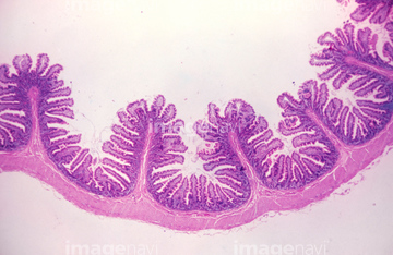
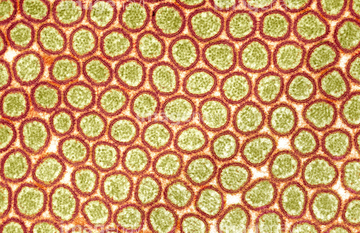
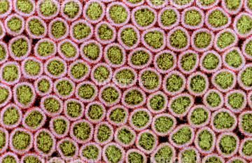
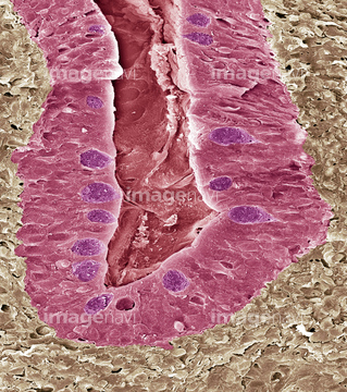
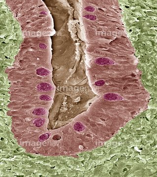
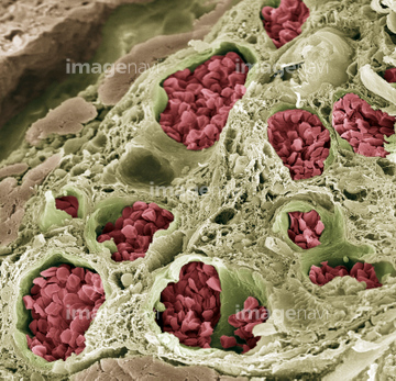
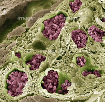
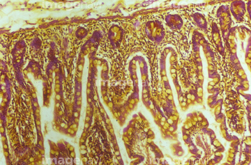
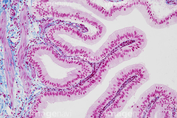
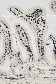
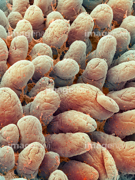
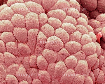
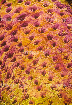
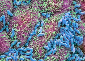
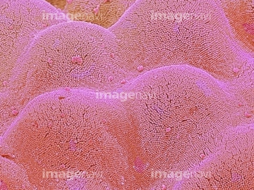
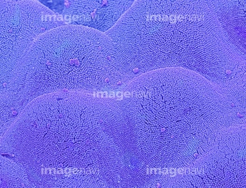
この検索結果には、Villi of the small intestine, SEM、Small intestine villus, TEM、Microvilli of the small intestine, TEM、Microvilli and a goblet cell、Small intestine lining, light micrograph、Intestinal lining, SEMなどが含まれています。

64225133

64225083
64206819

64225140
64196734
64055781
64049910
64152547
64075661
64055763
64049911
64021549
64021550
64021551
64008180
64225256
64251226
64251229
64224937
64225131
64021569
64021548
64021552
64021534
64150338
64021537
64021539
64021540
64021541
64021571
64061536
64021570
64088343
64209800
17202471
17202472
64132948
64132949
64147742
64225094
64021568
64262811
64262812
64262814
64262815
64021536
64247696
64234667
64234718
64213913
64213914
64213915
64021533
64077368
64021545
64056271

64133039
64223998
64223999
64002984
64002985
17255816
64190895
64060201
64060202
64225176
64116645
64114169
64225151
64225152
64114553
64114554
64021532
64021535
64021538

64146135

64146275
64114552
64224842
64221011
64264588
64264589
64090479
64090480
64196715
64073608
64074891
64076360
64076361
64225092
64021554
64056746
64021528
64021553
17251192
17251193
17251194
64087883
64209773
64209785
64209786
64211723
64211724
64211725
64224938
64132258
64132996
64132997
17201424
64021580
64114561
64021591
64021592


64104713
64104724
64104731
64104736
64060825
64060826
64008744
64008745
64008746
64008751
64234509
64234510
17201425
64077369
20536138
20536141
64042875
64197097
64066076
| 次ページ |
