HOME > 写真 > 科学・テクノロジー > 科学 > DNA・細胞
10,000件の写真素材が検索されました。
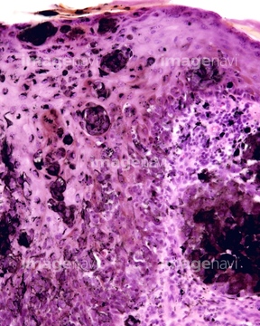
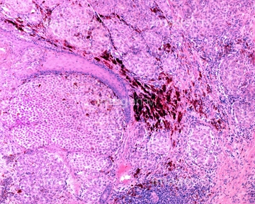
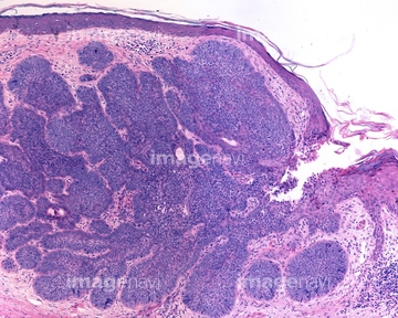
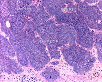
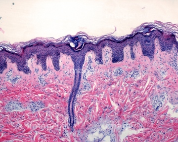
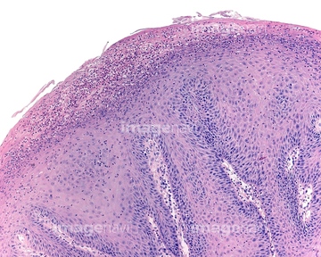
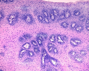
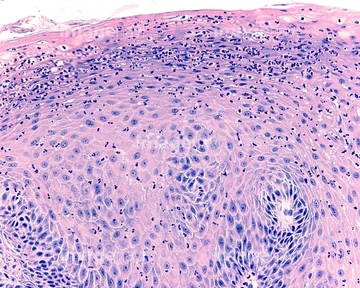
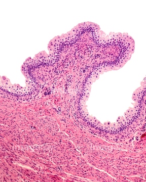
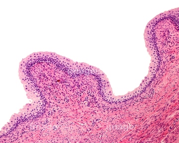
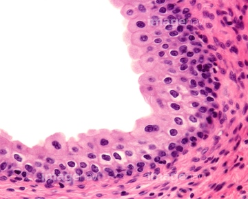
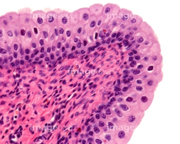
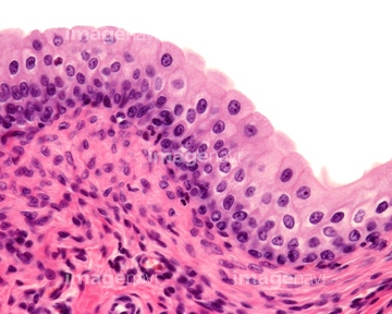
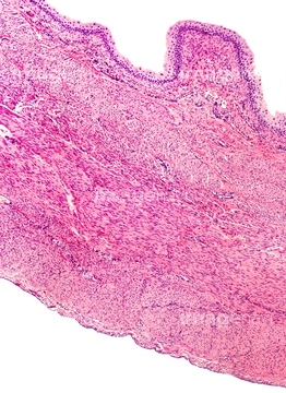
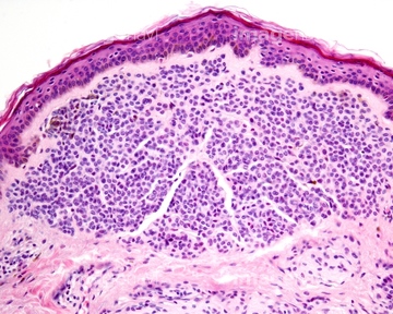
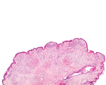
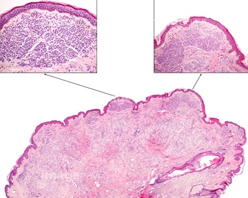
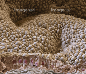
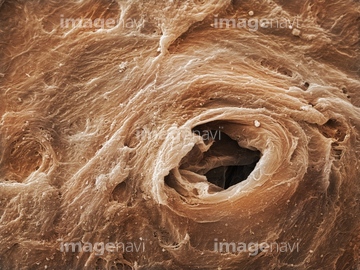
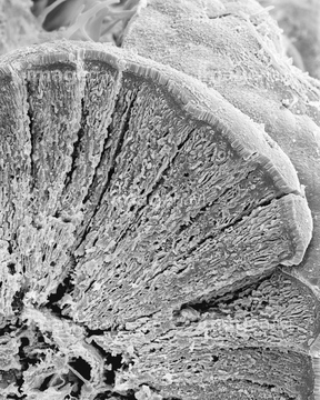
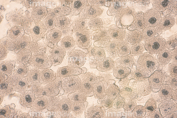
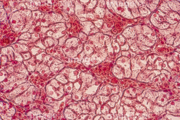
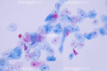
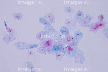
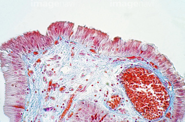
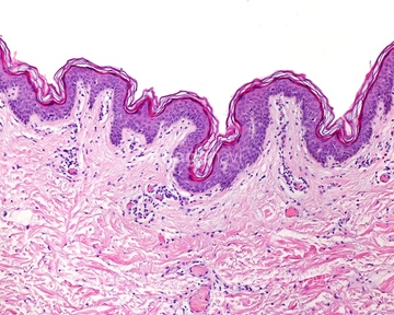
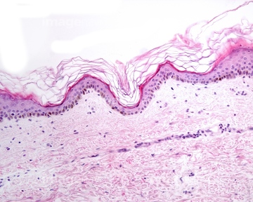
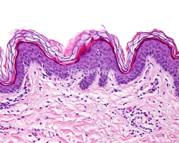
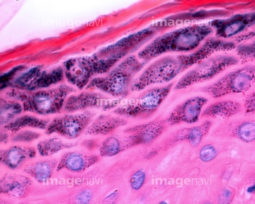
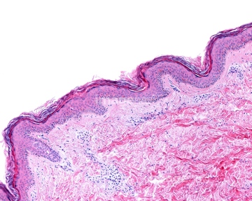
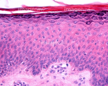
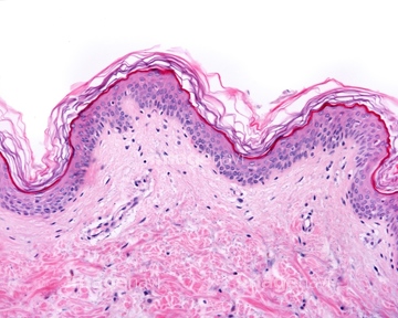
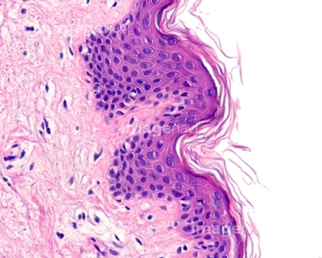
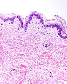
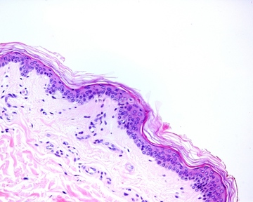
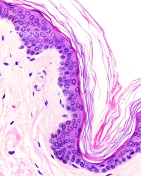
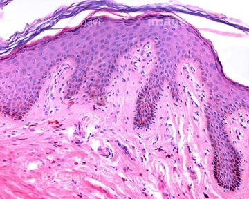
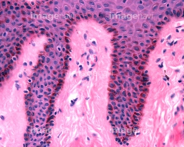
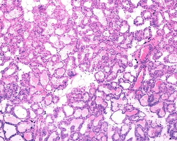
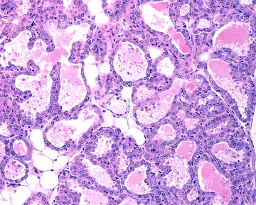
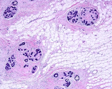
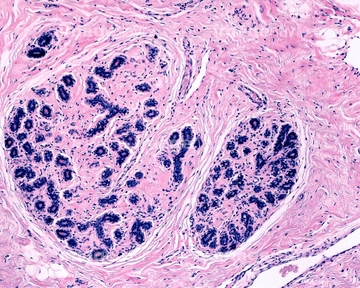
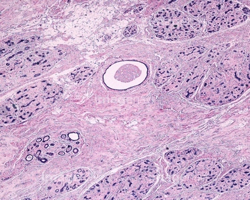
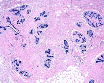
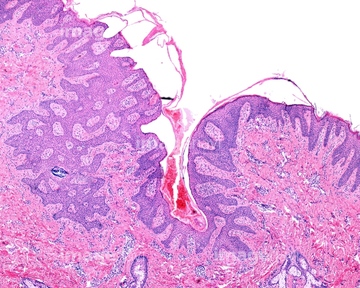
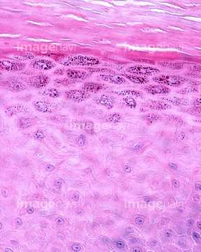
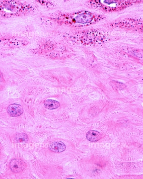
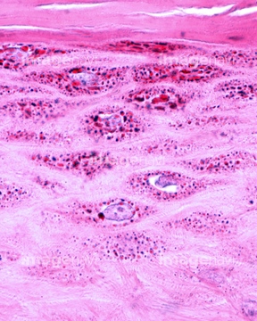
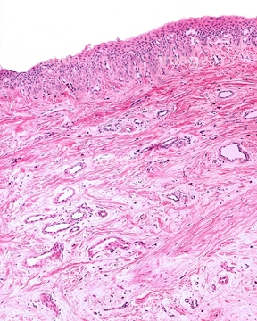
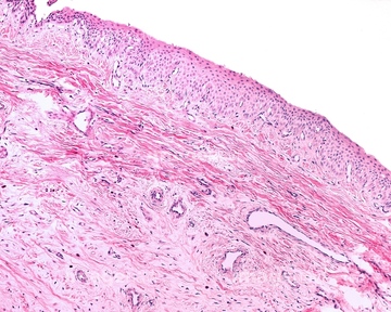
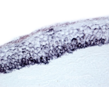
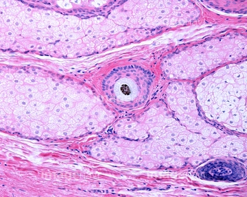
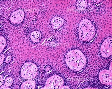
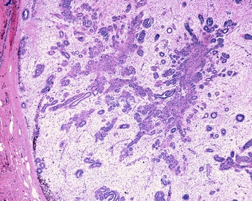
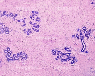
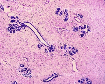
この検索結果には、Nevoepithelioma, light micrograph、Basal cell carcinoma, light micrograph、Fibroma of the skin, light micrograph、Soft palate papilloma, light micrograph、Urinary bladder, light micrograph、Wart, light micrographなどが含まれています。
64225158
64213665
64213672
64213773
64254942
64254947
64254950
64254958
64224841
64075613
64075614
64169977
64062293
64062294
64090469
64060069
64102361
64017970
64017971
64017982
64217238
64217243
64217244
64217246
64217254
64217258
64224950
64223922
64112127
64112128
64017969
30015123
64022286
64225199

64104713
64104724
64104731
64104736
20574390
64056754
64169967
64197456
64197460
64209512
64209513
64209514

64209515
64176896
64147075
64102368
64061550
64225200
64111448
64116976
17201561
64114169


64225083
64225131

64225133

64225140
64225151
64225152
64225259
64150338
64167101
64167102
64167104
64167105
64167108
64167110
64167113
64167114
64167115
64167116
64167117
64167118
64167119
64167121
64167122
64167123
64167124
64167127
64167128
64169711
64169714
64169715
64169725
64169734
64169949
64169950
64169961
64169962
64169978
64205381
64180278
64180279
64180280
64180282
64180295
64180296
64180297
64180298
64223998
64223999
64245076
64075579
64075580
17237231
17237232
20574313
20574346
20563355
20563356
64224892
64150218
64150226
64150227
64150228
64150268
64150270
64150280
64150294
64150323
| 次ページ |
