HOME > 写真 > 科学・テクノロジー > 科学 > DNA・細胞
10,000件の写真素材が検索されました。
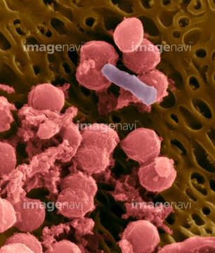
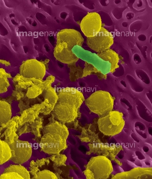
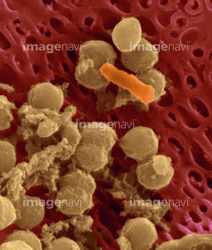
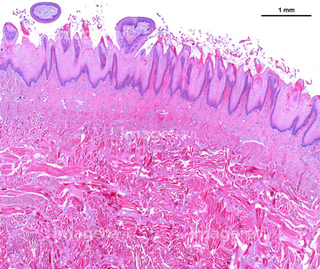
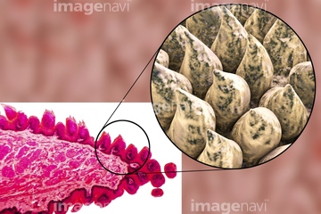
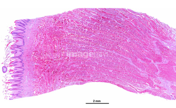
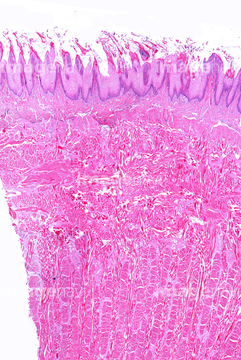
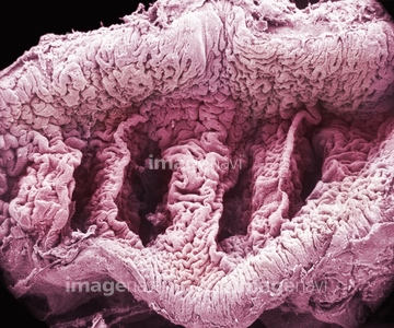
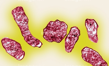
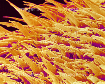
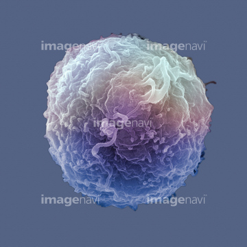
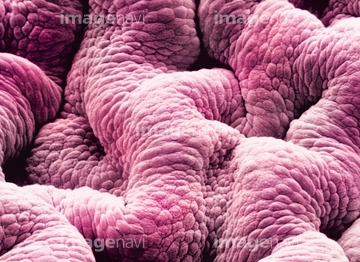
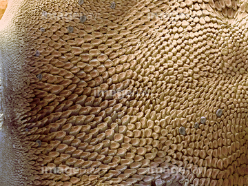
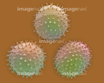
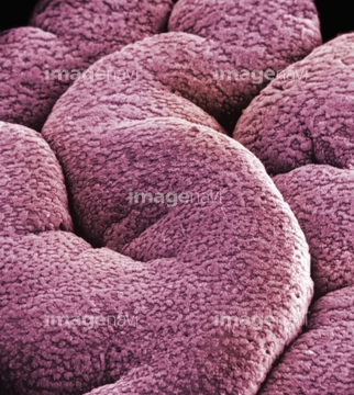
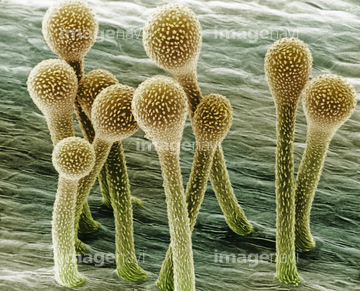
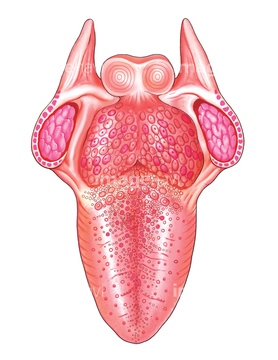
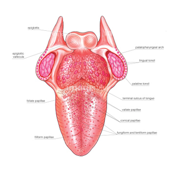
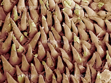
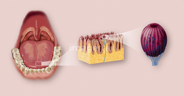
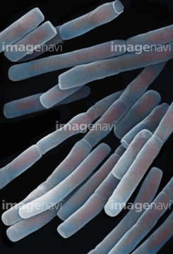
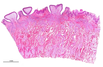
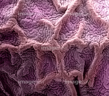
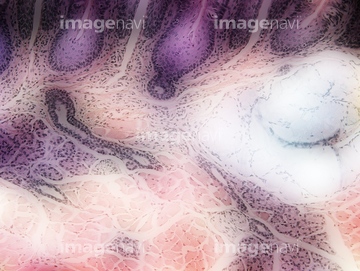
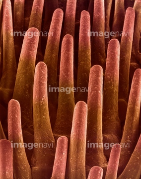
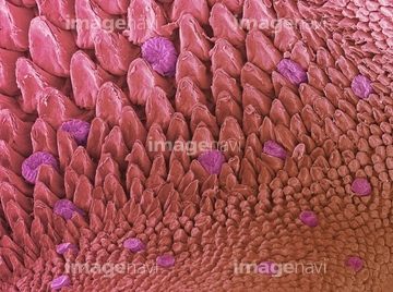
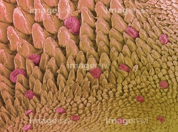
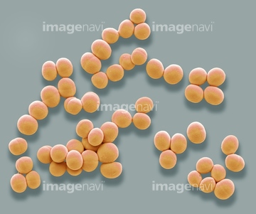
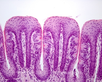
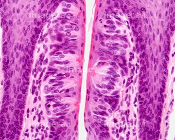
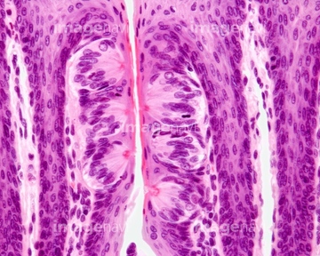
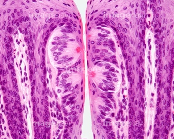
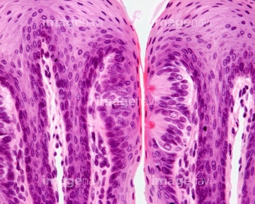
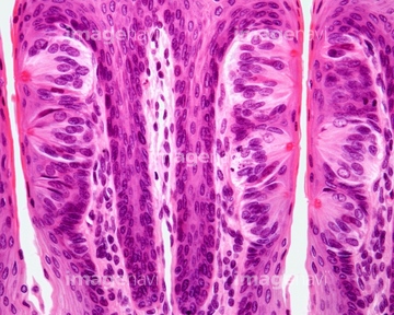
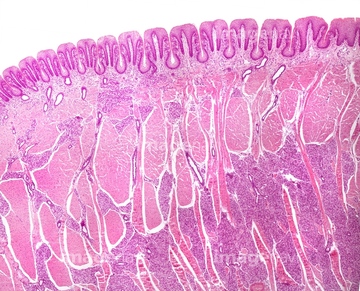
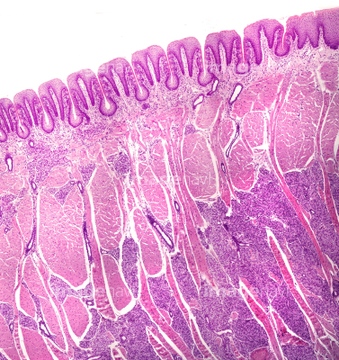
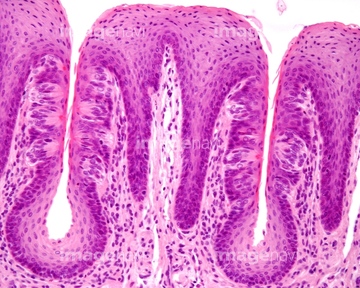
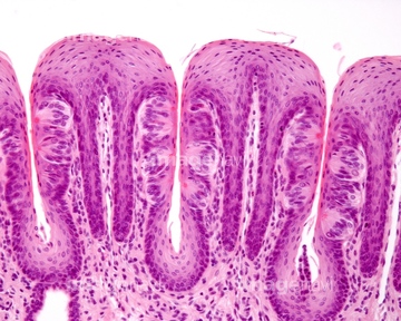
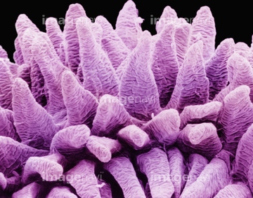
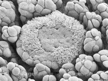
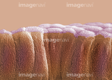
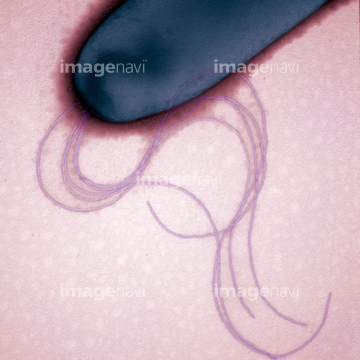
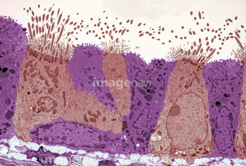
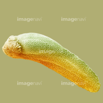
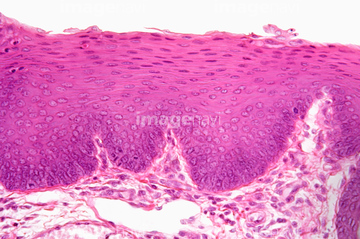
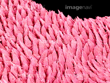
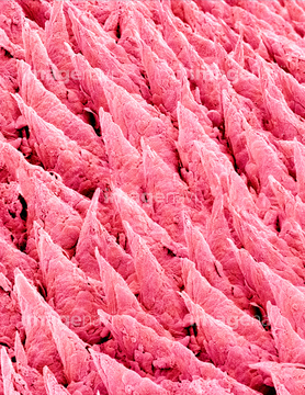
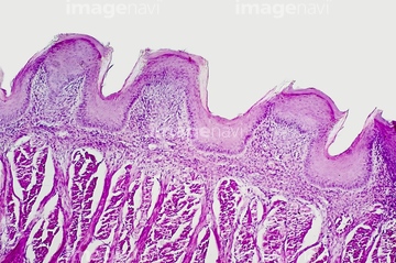
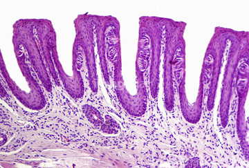
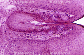
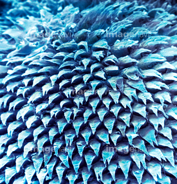
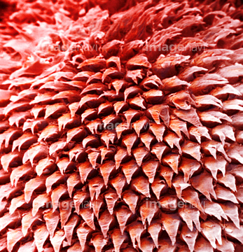
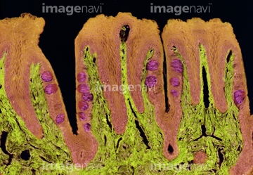
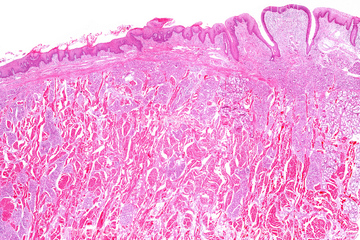
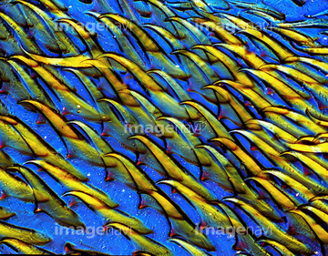
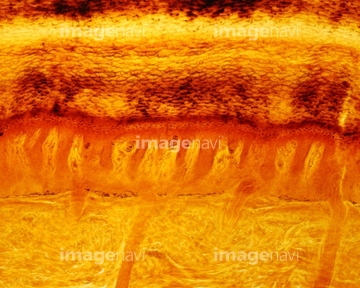
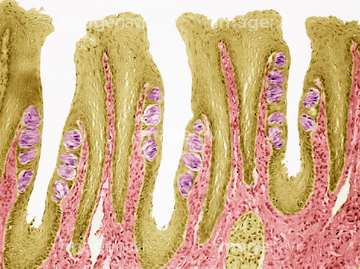
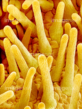
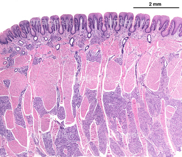
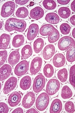
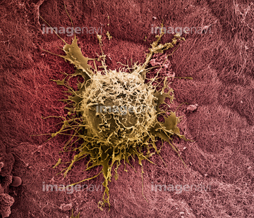
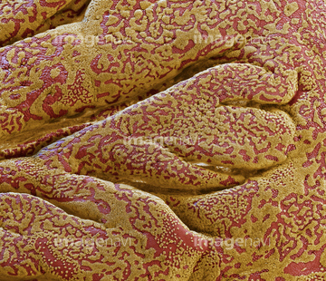
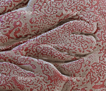
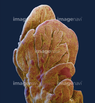
この検索結果には、Tongue anatomy, artwork、(Bacillus megaterium) Bacteria、Vallate papillae of human tongue, light micrograph、The Rabbit gallbladder、Tongue tissue, light micrograph、Mouse tongue surface, SEMなどが含まれています。
64225257
64225082
20523444
64035026
64035025
64225265
17261424
17261425
17261426
17261427
64021492
64021493
64115471
17255811
17255812
64254288
64254289
64225261
64115493
64115600
64225361
64010899

64010900
64010901
64010902
64011856
64011858
64011860
64011861
64011862
64056743

64225259
64225201
64180368
64035536
64035537

64115490
64071775
64071776
64035021
64050501
64224880

64225260
64139404
64151066
64137595
64137596
64137597
64137598
64214801
64214818
17281852
17281881
17281895
17246260
17246264
17246265
64115520
64115521
64115526
64115593
64115594
64115595
64225091
64225243
64224854
64225086
64225190
64114730
64225175
17283421
64225232
64225233
64115535
64115536
17283419
64151678
64151683
64263580
64263598

64225133
64213898
64213899
64213900
64213901
64213902
64225256
64115597
64225242
64225244
64225264
64235181
64235182
64225176
64115602
64115596
64224868
64224871
64225101
64225157

64225083
64224897
64225069
64225070
64225195
64225197
64010903
64010904
64010905
64006154
64115443
64265118
64265146
64265159
64006119
64006151
64006156
64011388

64114540
64114561
64073616
64073617
64161462
64010886
64010887
64010888
64055782
20532887
20532889
20532894
64047098
64123469
64123483
64123499
64236634
64236655
64150013
64235183
64220282
64008177
64006155
64199723
64199724
64199737
64199738
64199739
64199740
64199741
64199742
64199743
64005415
64005416
64005678
| 次ページ |
