HOME > 写真 > 科学・テクノロジー > 科学 > DNA・細胞
10,000件の写真素材が検索されました。
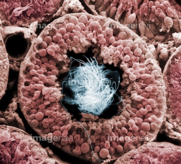
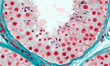
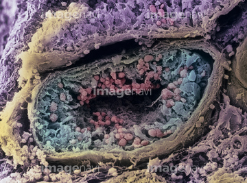
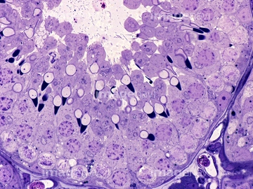
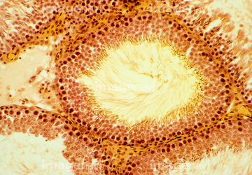
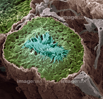
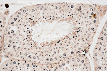
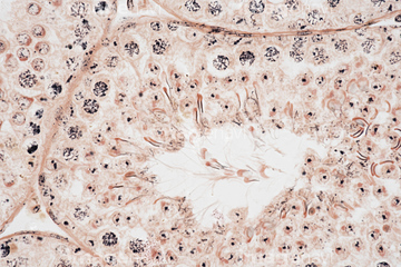
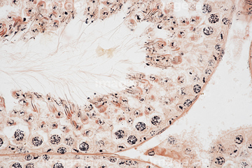
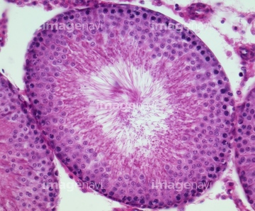
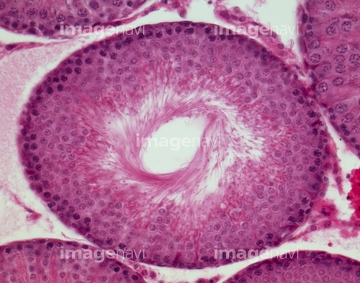
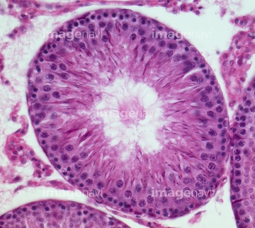
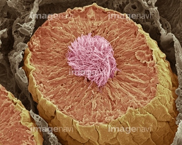
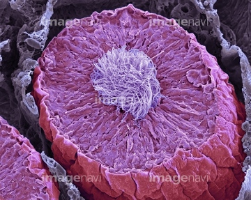
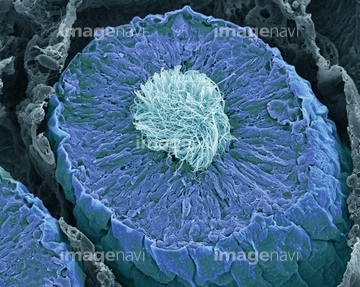
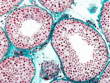
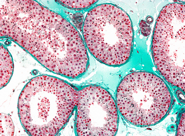
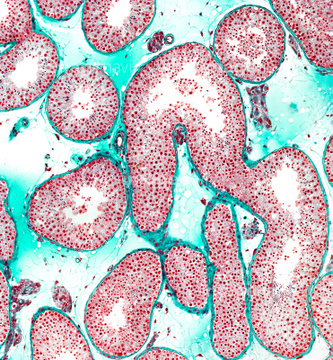
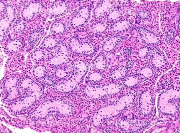
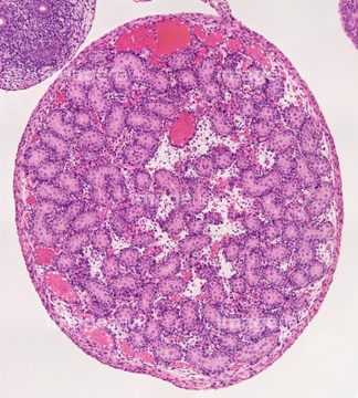
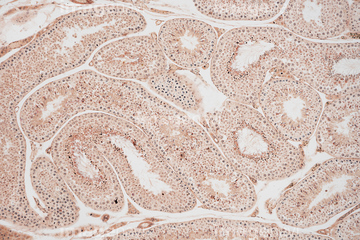
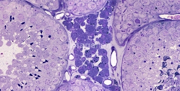
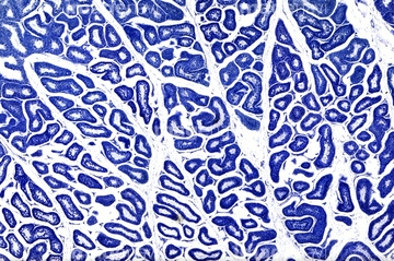
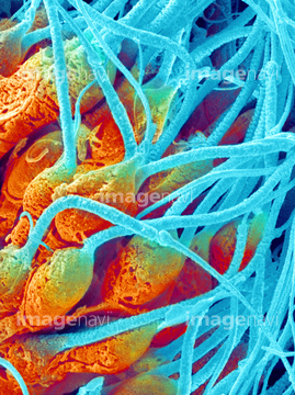
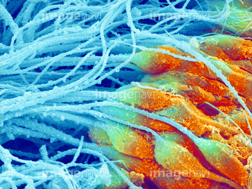
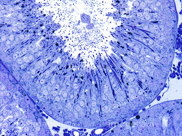
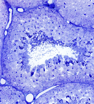
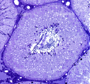
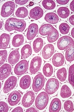
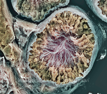
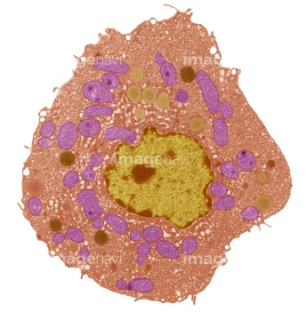
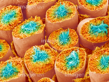
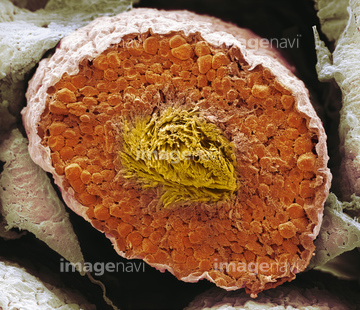
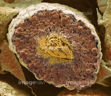
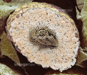
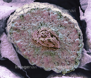
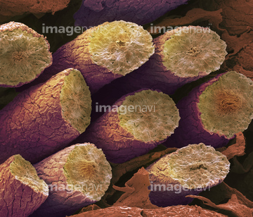
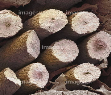
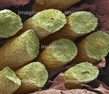
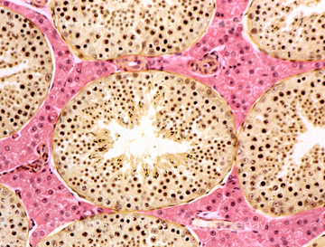
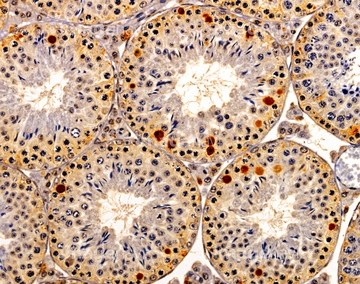
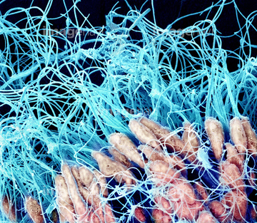
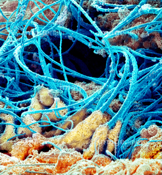
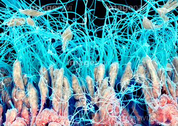
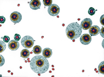
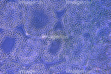
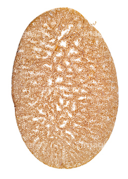
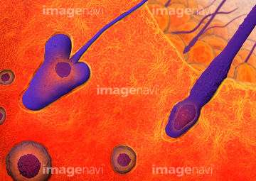
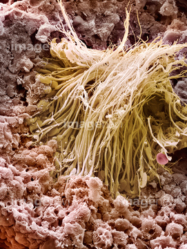
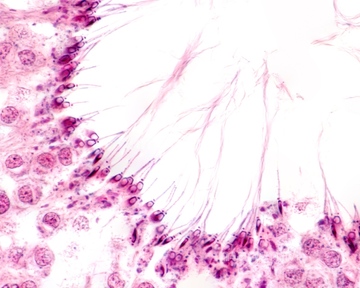
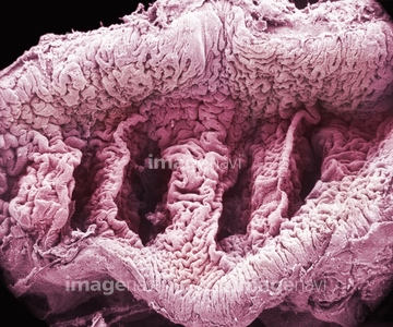
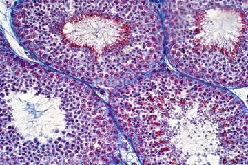
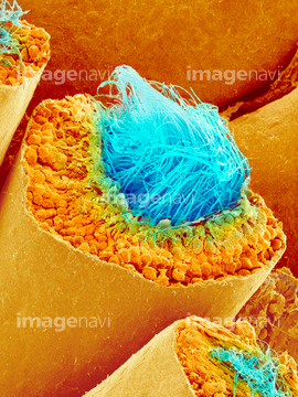
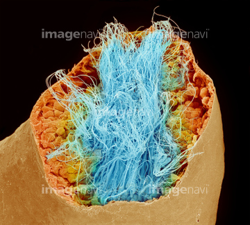
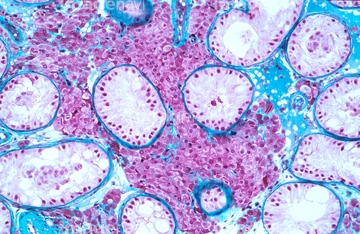
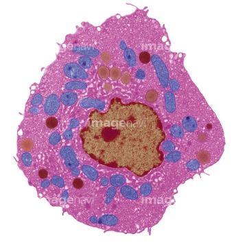
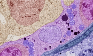
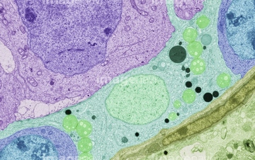
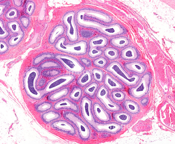
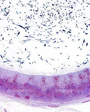
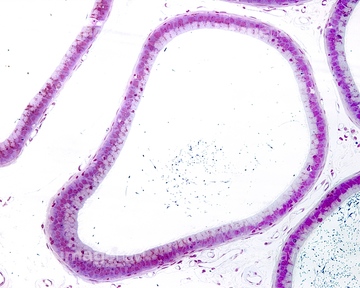
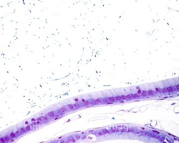
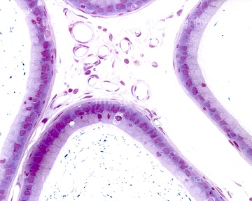
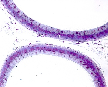
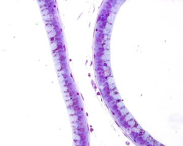
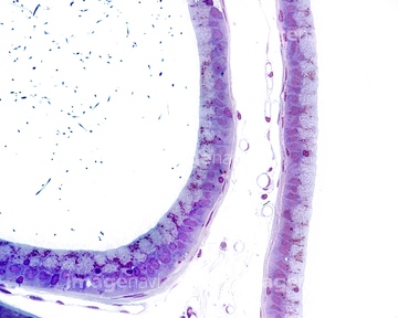
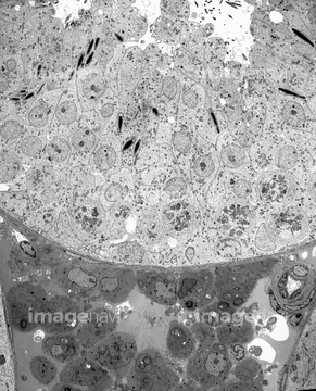
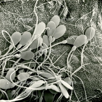
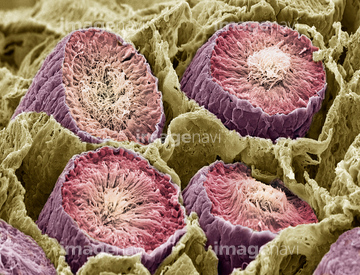
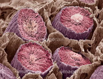
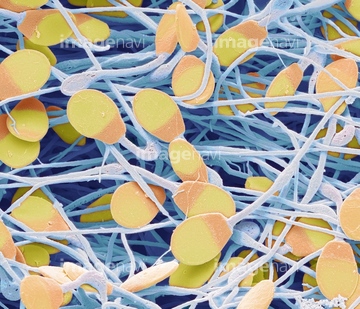
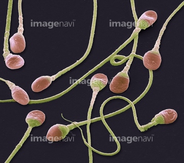
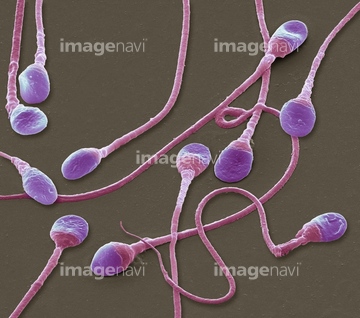
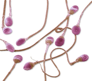
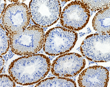
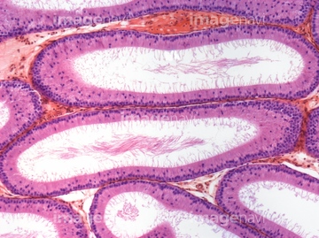
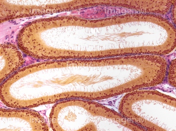
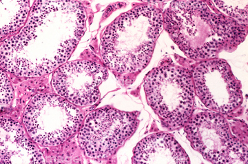
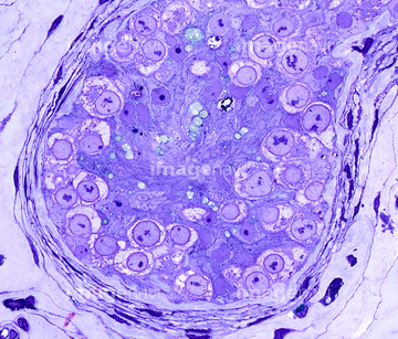
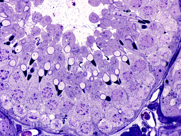
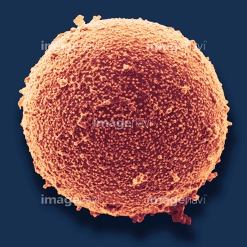
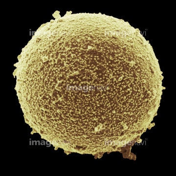
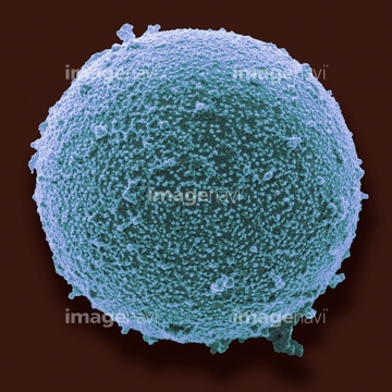
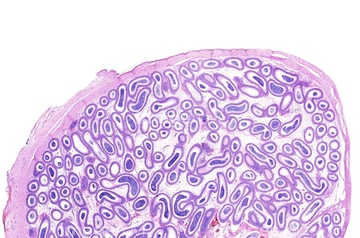
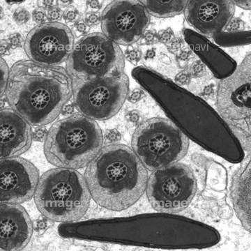
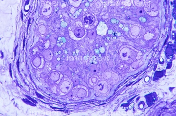
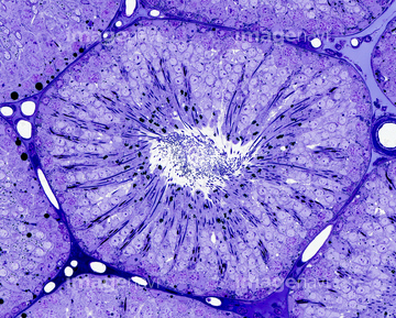
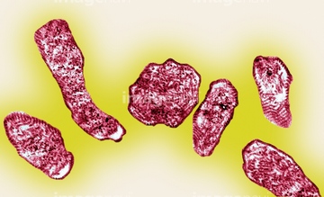
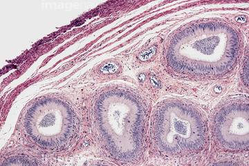
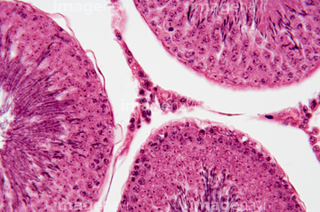
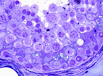
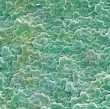
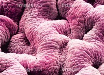
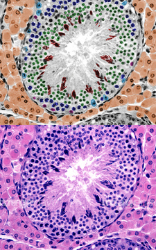
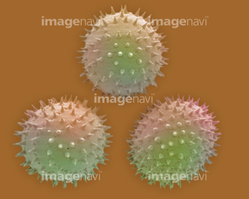
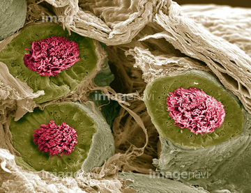
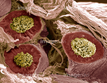
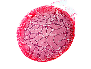
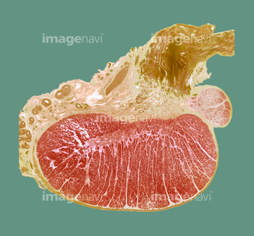
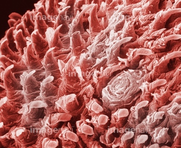
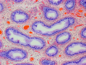
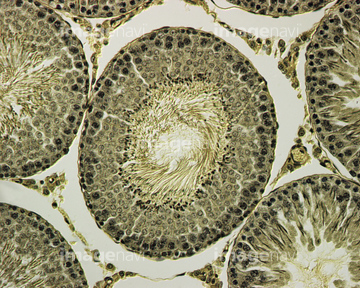
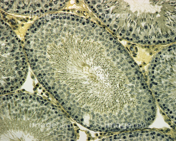
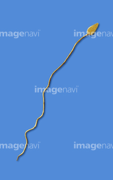
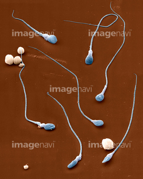
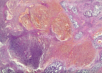
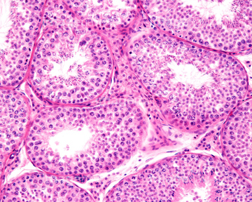
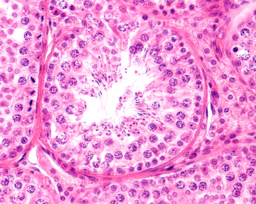
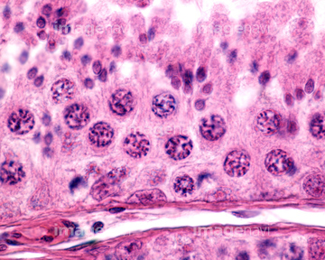
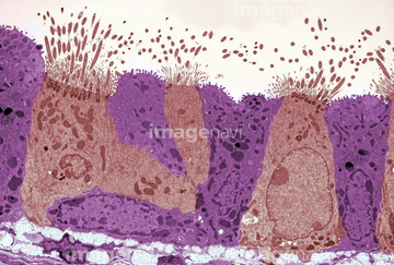
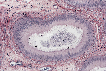
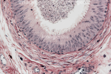
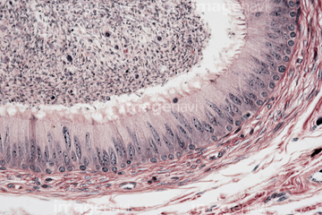
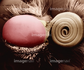
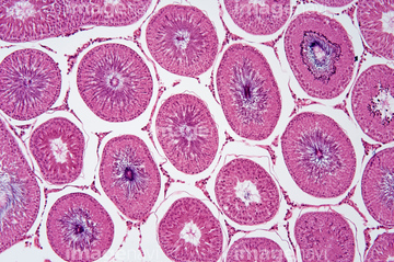
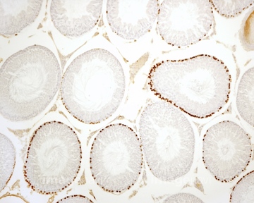
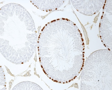
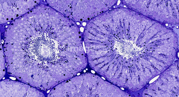
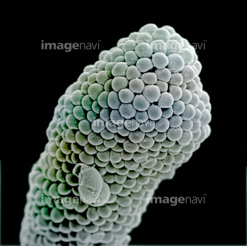
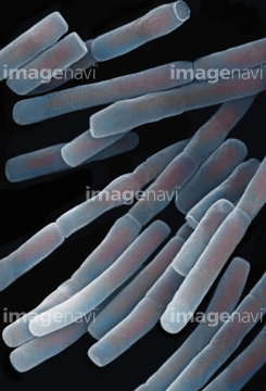
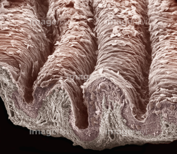
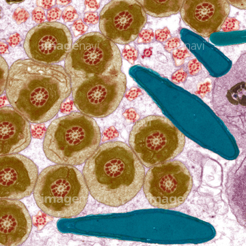
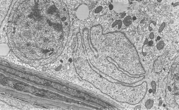
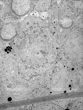
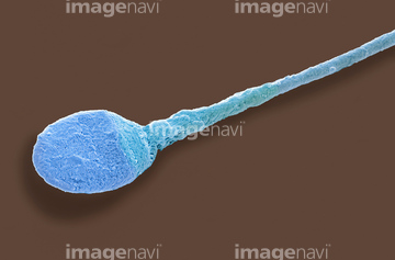
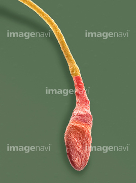
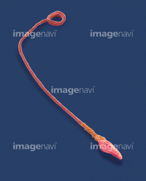
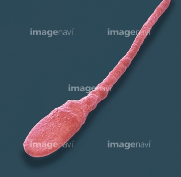
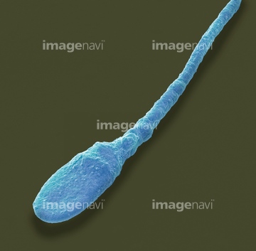
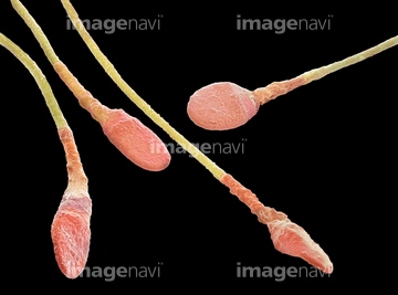
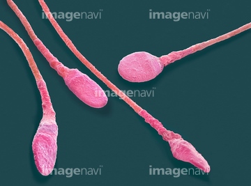
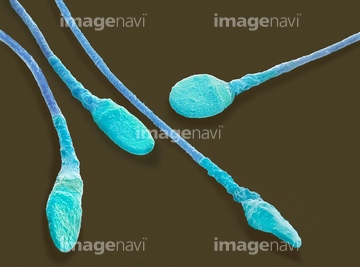
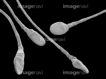
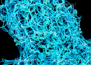
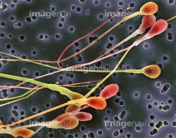
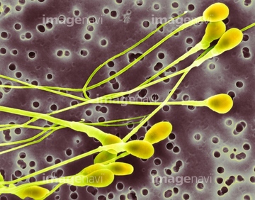
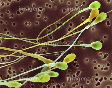
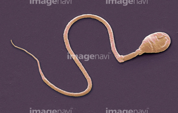
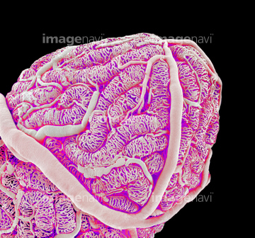
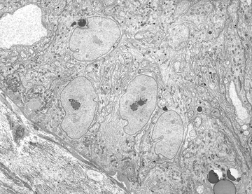
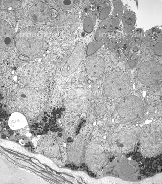
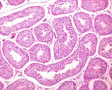
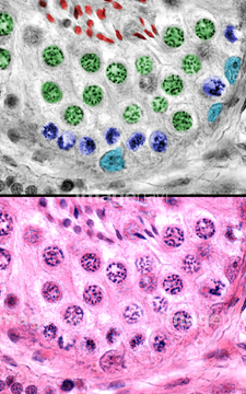
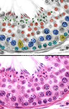
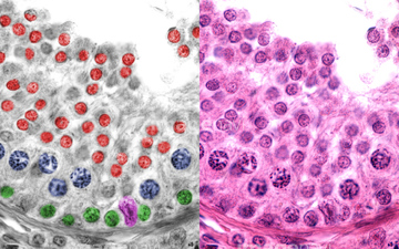
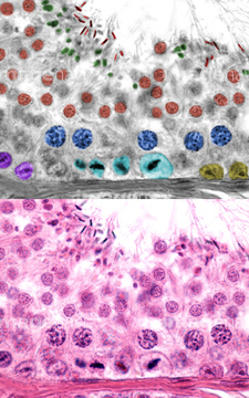
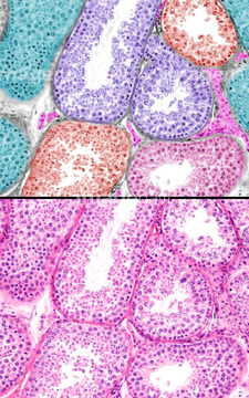
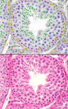
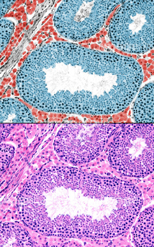
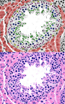
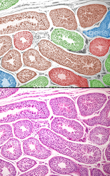
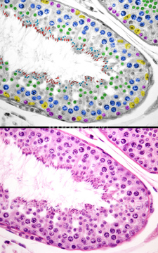
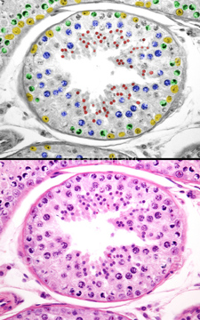
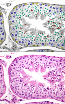
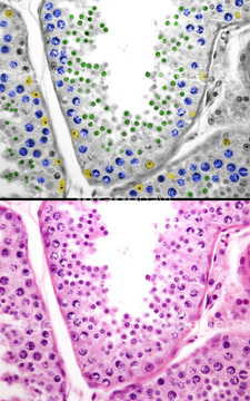
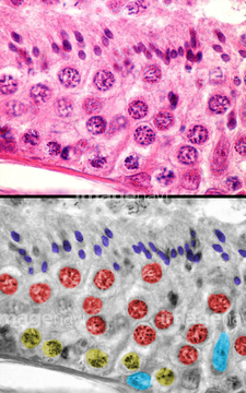
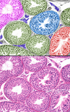
この検索結果には、Human sperm centriole, light micrograph、Distal end of the seminal vesicle、Human testis, light micrograph、Sperm production, SEM、Sertoli cell, TEM、Sertoli cells, TEMなどが含まれています。
64021709
64008178
20548464
20548475
20548500
64090473
64090477
64090478
64106072
64106095
64234644
64021721
64021722
64090151
64090165
64090173
64225361
64021716
64103901
64056750
64250181
64250183
64250186
64250187
64250192
64250194
64250199
64077495
64076289
64076290
64166161
64085235
64051054
17201432
64225261
20542510
64013157
64013158
64116656
64103884
64136245
64136255
64098234
64114543
64206745
64247700
64183414
17200056
17200057
17281862
20523341
20523351
20523376
64090501
64207216
64207257
64251234
64111600
64111608
64111633

64196721
64196722
64155033

64115471
64115493
64115600
64263112
64263113
64263114
64259998
64247692
64234967
64247438
64085315
64085325
64114544
64251238
64225259
64115490
64011397
64011398
64055677
64021723
64225257
64093983
64224856
64021803
64115596
64234968
64234969
64234970
64115535
64115536
17246260
17246264
17246265
64115520
64115526
64115594
64115595

64225091
64225243
64114730
64224880
64225175
64225201
64256395
64197109
64021811
64247686
64247693
17234574
17234576
17234581
64111621
64111623
64111639
64111640
64111641
64152610
64076288
64177496
64177497
64177498
64075153
64011825
64247689
64247718

| 次ページ |
