HOME > 写真 > 科学・テクノロジー > 科学 > DNA・細胞
10,000件の写真素材が検索されました。
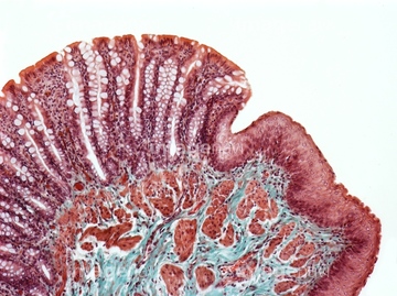
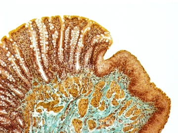
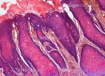
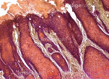
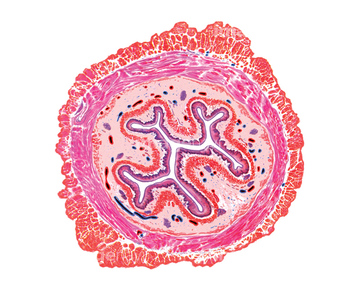
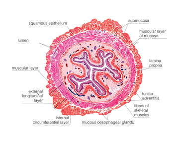
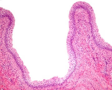
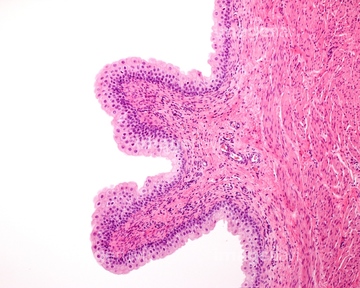
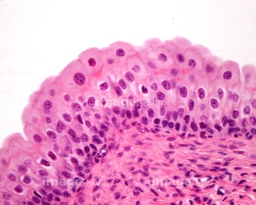
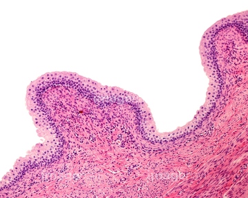
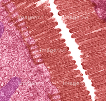
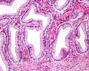
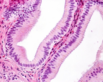
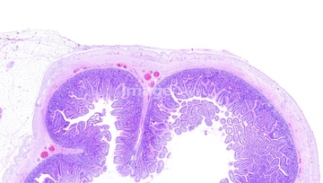
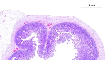
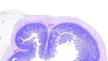
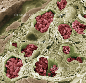
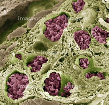
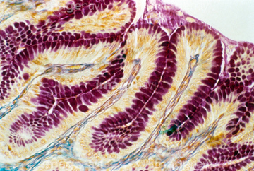
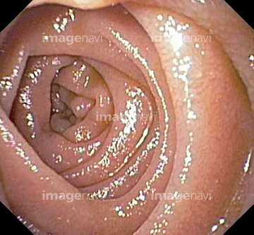
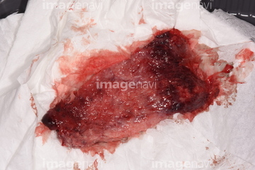
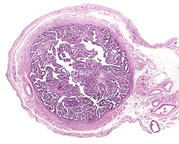
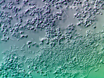
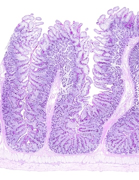
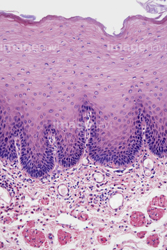
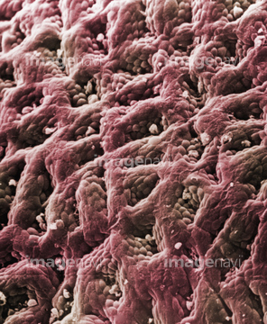
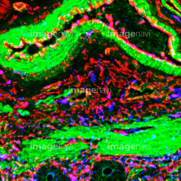
この検索結果には、バック版、細胞、Intestinal nerves, light micrograph、Oesophagus epithelium, SEM、Nervous system tumour, light micrograph、Stomach metaplasia, light micrographなどが含まれています。
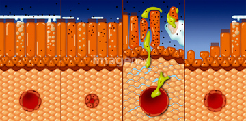
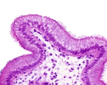
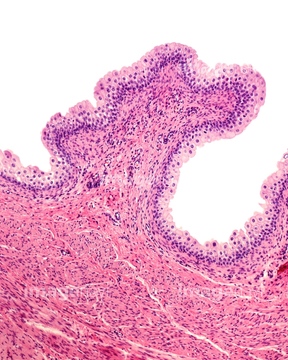
41243381
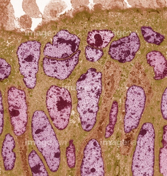
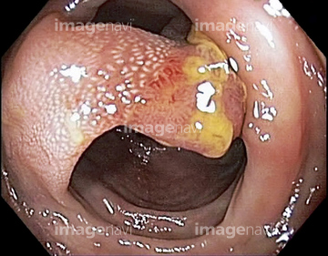
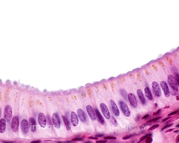
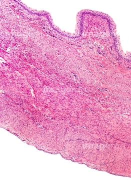
41243376
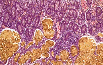
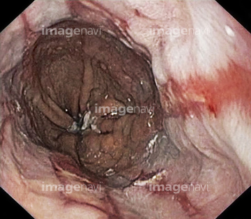
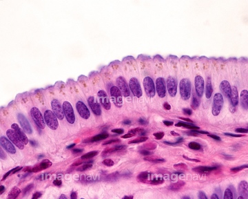
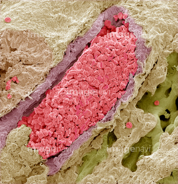
41243378
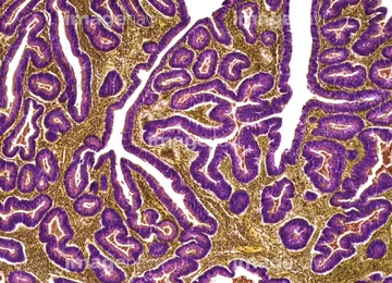
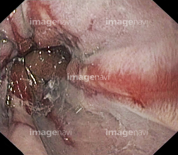
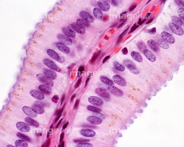
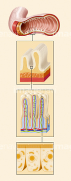
41243379
41243380
41243585
41243586
41243587
41243374
41243375
41243410
41243411
64050490
64009248
64009009
64009061
64010476
64021654
64021544
64205896
64205907
64207241
64207244
64176605
17200010
17200011
64071833
64071834
64010379
64003085
64172956
64212970

64212981

64213006
64155646
64155653
64155666
64155671
64021542
64021523
64021524
17201424
17201435
17200012
17200013
17200049
17200050
64088312
64094080
64092857
64143021
64143022
64197456
64200637
64200650
64177447
64216989
64216990
64056751
64060220
64045958
64050457
64254349
64225259
64225256
64225260
64008993
64010681
64011070
64159057
64184302
64184329
64176606
64176621
64176637
64176638
17202471
17202472
20544104

64146135

64146275
64161259
64161268
64197478
64235010
64221011
64213913
64213914
64213915
64008746
64008751
64071988
64071989
64072028

64072029
64073274
64073275
64073276
64055754
64170387
64170388
64170389
64170390
64170393
64170394
64170395
64170396
64170397
64265597
64003075
64022385
64180234
64162986
64162987
64071773
64071774
64071877
64071878
64071879
64071880
64071882
64071883
64071886
64071887
64071893
64055683
64143023
64155657
64155701
64045605
64050497
64013522
64018941
64136447
64206819
64235009
64225092
64008158
64008744
| 次ページ |
