HOME > 写真 > 科学・テクノロジー > 科学 > DNA・細胞
10,000件の写真素材が検索されました。
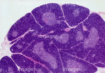
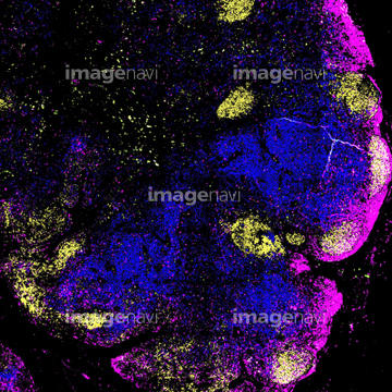
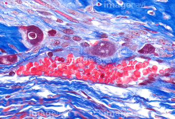
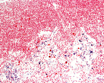
この検索結果には、Thymus gland, light micrograph、Lymph node, fluorescent light micrograph、Blood vessel, light micrograph、Lymph node macrophages, light micrograph、Venule, light micrograph、Red blood cellなどが含まれています。
64224953
64068306
64205382
64206772
64206771
64105458
64090502
64106064
64105464
64234835
64234836
64234837
64090464
64090467
64073294
64065599
64065600
64066067
64065592
64225293
64065613
64060819
64060820
64225130
64115571
64224954

64066069

64225172
64115600
64150208
64064560
64251245
64220925
64115471
64063053
64063054


64132310
64132311
64132312
64163250
64235123
64235125
64235126
64235128
64234489
64258373
64225179
64150210
64150278
64063025
64090152
64235008
64201907
64055761
64055762
64258069
64258070
64258072
64258078
64258079
64224851
64078928
64224951
64225232
64225233
64207935
64207936
64225198
64132607
64132609
64132610
64132611
64245184
64245187
64114552
64114561

64146135

64146275
64224945
64256627
64256631
64256632
64257061
64257062
64116674
64174621
64201881
64244165
64063008
64063050
64115493
64114526
64114529
64115581
64082463
64114522
64114523
64114524
64114540
64114549
64114570
64114571
17235958
64208213
64213924
64224932
64224950
64114521
64072732
64072767
64072770
64072773
64205881
64065997
64204230
64204232
64209749
64209762
64209763
64209791
| 次ページ |
