HOME > 写真 > 科学・テクノロジー > 科学 > DNA・細胞
10,000件の写真素材が検索されました。
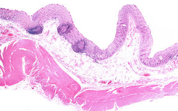
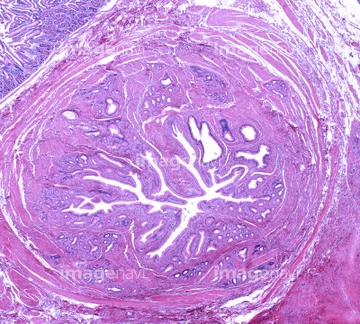
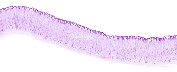
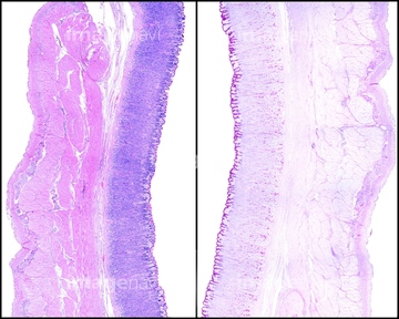
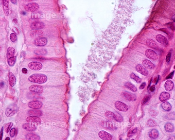
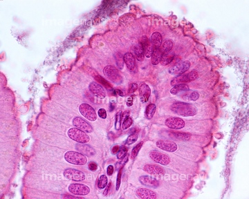
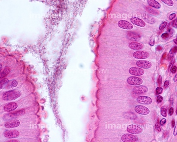
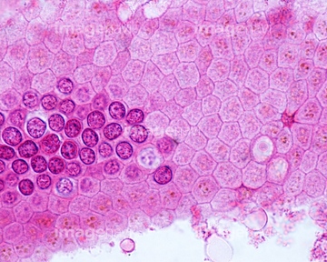
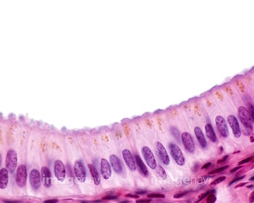
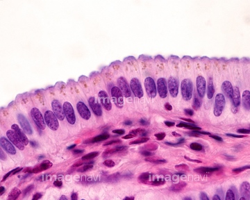
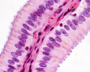
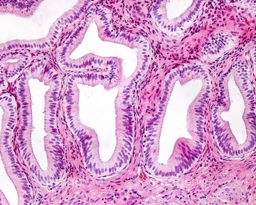
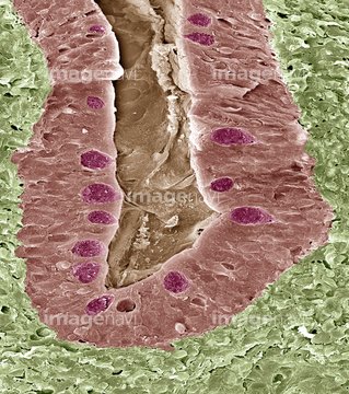
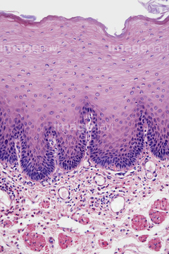
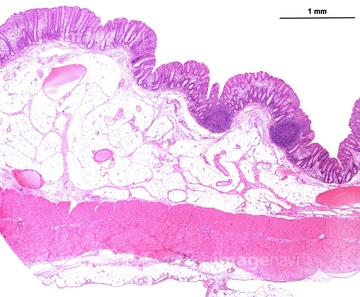
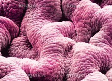
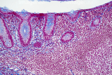
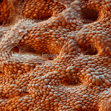
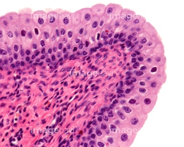
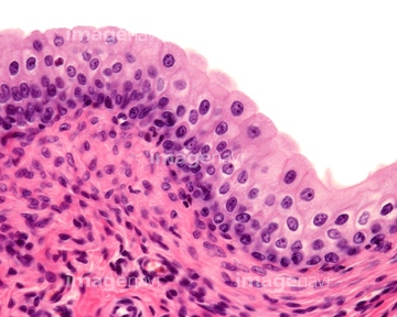
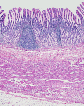
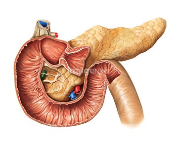
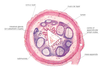
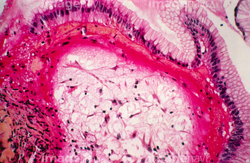
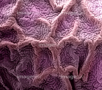
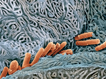
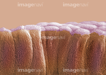
この検索結果には、Duodenum, light micrograph、Stomach lining, light micrograph、Colon lining, light micrograph、Large bowel, light micrograph、LM of a section through the stomach wall、Structure of intestinal tract, artworkなどが含まれています。
64225192
64225131

64084027
64090172
64071833
64071834
64071876
64071877
64071886
64071887
64071892
64071893
64197478
64065178
64071850
64071851
64055692
64055693
64245220
64021523
64021524
64225261
64055753
64085261
64085267
64196715
64196738
64197479
64197486
64197489
64213913
64213914
64213915
64071988
64071989
64250037

64225140
64225183

64146135

64146275
64221011
64196735
64008159
64176856
64176857
64176858
64225092
64008744
64008745
64008746
64008751
64235009
64235010
64066084
64225182

64206746
64206747
64225259
64234280
64056745
64197456
64234838
64071874
64071875
64152547
64136447
17202471
17202472
64190237
64215656

64225083

64225133
64082492
64073993
64074822
64225189
64115571
64225190

64008158
64234509
64234510
64235008
64029976
64224890
64072028

64072029
64225260
20507330
20507334
64082491
64073996
64011433
64011434
64011435
64011436
20542530
20544099
64196734
64199159
64206819
64234842
64213868

64213869
64073272
64073273
64073274
64073275
64073276
64106069
64114169
64115471
64224897
64245072
64225069
64225070
64225088
64225129
64225130
64225132
64225151
64225152
64225172
64225197
64225198
64225201
64225202
64225361
64225001
64225159
64021511
64021512
64225176
| 次ページ |
